the midline sternotomy in the stable patient with imaging

Pacing Options in the Adult Patient with Congenital Heart Disease - part 1 pptx
Ngày tải lên :
14/08/2014, 07:20
... utilizing their individual expertise This book outlines the principles of dealing with such patients including the preferences of techniques and the hardware available The text is divided into ... Acquisitions: Gina Almond Development: Beckie Brand Set in 10/13 Palatino by Newgen Imaging Systems (P) Ltd., Chennai, India Printed and bound in Singapore by COS Printers Pte Ltd For further information ... trunk into the superior vena cava Right: A left azygous (hemiazygous) vein drains blood from the lower trunk into a left superior vena cava and from there into the right atrium via the coronary sinus...
- 12
- 335
- 0

Pacing Options in the Adult Patient with Congenital Heart Disease - part 2 potx
Ngày tải lên :
14/08/2014, 07:20
... invaginate the vein into the subclavian vein The torn cephalic vein then produces a choking collar around the leads preventing positioning of the atrial lead (Figure 2.5) In this situation, the ... vein retained guide wire technique Left: Two leads are passed along an intact cephalic vein Right: The cephalic vein is torn with pushing of the second lead into the subclavian vein The vein ... introducer is passed along the guide wire into the cephalic vein Right: The dilator of the introducer is removed leaving the guide wire The lead is then inserted parallel to the guide wire technique,...
- 15
- 377
- 0

Pacing Options in the Adult Patient with Congenital Heart Disease - part 3 docx
Ngày tải lên :
14/08/2014, 07:20
... but rather proceed retrograde towards the arm in the axillary vein or up into the neck in the internal jugular vein (Figure 7.4) Although this can usually be prevented by not peeling the introducer ... through the annuloplasty ring to the apex of the right ventricle The other lead (white arrow) lies in the body of the right atrium The operation of the Locator® produces only a small change in the ... chambers The advantage to the Locator® is that the curve can be created from the straight position without removing the stylet In the center, the preformed atrial J stylet allows the lead to enter the...
- 15
- 406
- 0

Pacing Options in the Adult Patient with Congenital Heart Disease - part 4 pdf
Ngày tải lên :
14/08/2014, 07:20
... cava will result in marked difficulties in trying to achieve left ventricular pacing The incidence of this abnormality draining into the coronary sinus is about 3–5% of patients with structurally ... biventricular pacing An enlarged thebesian valve within the coronary sinus may obstruct the ostium making lead positioning difficult or impossible 39 40 Chapter 11 In patients with D-transposition of the great ... outside the vein in the pacemaker pocket has also been suggested The lead is secured to the tissues via the lead collar, using slowly absorbable suture material The lead can then be drawn into the intravascular...
- 15
- 344
- 0

Pacing Options in the Adult Patient with Congenital Heart Disease - part 5 doc
Ngày tải lên :
14/08/2014, 07:20
... ventricular lead passing inferior without a bend as if into the floor of the right atrium and fits nicely with the accompanying schematic Depending on its position in the ventricle, the lead tip may ... 12-lead ECG from the same patient demonstrating dual chamber pacing with an inferior axis and the characteristic tall R waves from V2 to V6 side of each other whereas in the normal, the right lies ... question now being asked is whether left ventricular or biventricular pacing should be considered in the young patient with congenital atrioventricular block In a highly symptomatic patient with an...
- 15
- 301
- 0

Pacing Options in the Adult Patient with Congenital Heart Disease - part 6 pot
Ngày tải lên :
14/08/2014, 07:20
... show the ventricular lead emerging from the coronary sinus looping in the right atrium and then passing to the floor of the right ventricle The atrial lead (At) lies against the lateral wall of the ... reported with the incidence falling with advancing age, but still as high as 20% in the elderly [148] Atrial septal defects are also one of the most common congenital defects occurring in adults with ... to show the ventricular lead emerging from the coronary sinus, looping in the large right atrium and passing through the tricuspid valve to the floor of the right ventricle (white arrow) The atrial...
- 15
- 384
- 0

Pacing Options in the Adult Patient with Congenital Heart Disease - part 7 potx
Ngày tải lên :
14/08/2014, 07:20
... (RAO) showing dual chamber pacing in a patient with Ebstein’s anomaly The passive-fixation atrial lead lies in the atrial appendage The passive-fixation right ventricular lead lies in the atrialized ... showing dual chamber pacing in a patient with the Mustard procedure In all views, the upper active-fixation lead is attached to the roof of the left atrium, but unlike in Figures 20.3 and 20.4, the ... pacing with a right bundle branch block appearance and the axis will depend on where the lead lies For instance, if the lead lies low in the heart such as in the middle or lateral cardiac vein,...
- 15
- 403
- 0

Pacing Options in the Adult Patient with Congenital Heart Disease - part 8 pps
Ngày tải lên :
14/08/2014, 07:20
... showing dual chamber pacing in a patient with the Mustard procedure In the PA view, active-fixation leads are attached to the roof of the left atrium and lateral wall of the left ventricle In the ... operatively the coronary sinus may not drain directly into the right atrium The surgeon, in order to protect the conducting system may position the prosthetic tricuspid valve, so that the coronary sinus ... Resting 12-lead ECG from the same patient demonstrating dual chamber pacing with a right bundle branch block indicating left ventricular pacing There is a right axis deviation suggesting that the...
- 15
- 359
- 0

Pacing Options in the Adult Patient with Congenital Heart Disease - part 9 doc
Ngày tải lên :
14/08/2014, 07:20
... Prejean CA Pacing in the middle cardiac vein in a patient with tricuspid prosthesis PACE 2002; 25: 243–244 73 Bos HS, Pop GAM, Stel EA et al Dual site coronary sinus pacing in a patient with an artificial ... portion of the single ventricle The two epimyocardial leads are on top of each other in the PA view which has been highlighted with a box In the L Lat view, a black arrow points to the two epimyocardial ... puncturing the intra-atrial tunnel with a trans-septal needle [63, 253] There have been successful cases of transvenous lead positioning in patients with univentricular hearts, who have not had the...
- 15
- 348
- 0

Pacing Options in the Adult Patient with Congenital Heart Disease - part 10 pptx
Ngày tải lên :
14/08/2014, 07:20
... et al Permanent pacing in patients with univentricular heart (Abstract) PACE 1994; 17: 806 251 Blackburn MEC, Gibbs JL Ventricular pacing from the coronary sinus in a patient with a Fontan circulation ... tract, 103 innominate vein, internal jugular approach, limitation, interrupted inferior vena cava, 41 intra-atrial baffle, 40, 89, 91 repair, 41 introducers, funnel introducer, 6, second introducer, ... al Transvenous ventricular pacing options in Ebstein’s anomaly Indian Heart J 1998; 50: 565–568 201 Andersen C, Oxhøj H, Justensen P Pacing in a patient with Ebstein’s anomaly PACE 1989; 12: 1586–1588...
- 11
- 455
- 0
Assessment and Management of the Elderly Patient with Multiple Sclerosis pot
Ngày tải lên :
22/03/2014, 14:20
... spine or into limbs with neck flexion (Lhermitte’s sign), dysesthetic pain, back pain, visceral pain and pain secondary to muscle spasms.17 In the older population, these pain conditions may interact ... difficulties with abstract reasoning, word finding, and Healthy Aging & Clinical Care in the Elderly 2012:4 Elderly patient with multiple sclerosis certain visuospatial functions, and slowness in the ... 237 patients from 39 centers in the US and Canada aged 18–70 years with clinically defined MS They found that patients on the dalfampridine showed a 25% improvement in walking speed based on the...
- 11
- 508
- 0
Báo cáo y học: " Mitral valve replacement via right thoracotomy approach for prevention of mediastinitis in a female patient with long-term uncontrolled diabetes mellitus: a case report" potx
Ngày tải lên :
10/08/2014, 09:22
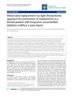
... http://www.cardiothoracicsurgery.org/content/5/1/38 conventional sternotomy would raise the risk of mediastinitis, the right thoracotomy approach was chosen to prevent mediastinitis by avoiding the splitting the sternum Through the right 4th ... of clinical data, the patient was able to walk without any complaints, indicating NYHA classification of 1/4 Postoperative infectious signs were not recognized (Figure 1a and 1b), and the patient ... mid-line full sternotomy was counter-indicated in this patient In view of the risk factors, a surgical approach considered more suitable for patients at risk for mediastinitis was selected During...
- 3
- 232
- 0

Báo cáo y học: "Unstable angina early after aortic valve replacement surgery in a female patient with normal coronary arteries preoperatively – a case report" pot
Ngày tải lên :
10/08/2014, 10:20
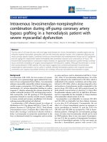
... [2]; balloon inflation in the proximal parts of the vessels; turbulence that causes coronary artery intimal injury that might explain the lesions often found distant to the adherence of the cannulation ... contributions SG was the main author and wrote the article CKN was the surgical consultant, was involved in data collection and revised the manuscript CS was the surgical consultant was involved in data collection ... surgery The procedure was performed using mini -sternotomy with a single 2-stage venous cannula and normothermic Figure left coronary artery Preoperative coronary angiography (RAO view) showing the...
- 4
- 346
- 0
Báo cáo y học: "Intravenous levosimendan-norepinephrine combination during off-pump coronary artery bypass grafting in a hemodialysis patient with severe myocardial dysfunctio" pps
Ngày tải lên :
10/08/2014, 10:20
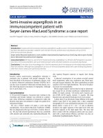
... 53 on the right) Despite the need to gradually increase norepinephrine dose to 0.15 μg/kg/min, in order to maintain MAP >60 mmHg, CI and SvO2 continued to rise, while CVP, PA and PAOP declined ... stability in this case In conclusion, this case report suggests that combined levosimedan/norepinephrine IV infusion is a reasonable inotropic support option in patients with heart failure and ongoing ... (albumin plasma level was 3.1 mg/dL in this case) In addition, the use of a low levosimendan dose, the combination with norepinephrine and close monitoring, in an attempt to avoid or promptly...
- 4
- 386
- 0

báo cáo khoa học: " Hypercalcemia and huge splenomegaly presenting in an elderly patient with B-cell non-Hodgkin’s lymphoma: a case report" potx
Ngày tải lên :
11/08/2014, 02:21
... other hematological malignancies, Figure An abdominal CT scan of the patient during the first hospital admission Figure PTHrP immunostaining Table Laboratory data of the patient on admission Patient ... was a major contributor in the writing of the manuscript and coordinated all members of the group AA performed splenectomy on our patient ED performed the gastrointestinal work up EF undertook ... [6,8-15] Table shows the clinical and laboratory data of 10 such patients, including ours As shown in Table 2, the hypercalcemia was severe and life-threatening and immediate therapeutic modalities...
- 4
- 254
- 0

Báo cáo y học: "Vertebral hyperostosis, ankylosed vertebral fracture and atlantoaxial rotatory subluxation in an elderly patient with" ppsx
Ngày tải lên :
11/08/2014, 10:22
... introduced to relieve pain and at the same time is capable of integrating into the bony matrix [11] Conclusion In the light of our findings, we believe that the degree of osteoporosis of the adjacent vertebral ... extremities Bowel and bladder sphincter control were all within normal limits No complaint of pain in her pelvis Examining the rest of her joints revealed nothing of significance Her height, weight ... hyperostosis (DISH) is an ossifying, non-inflammatory, non-erosive enthesopathy favouring the dorsal spine but sparing the sacroiliac joints DISH affects 3–6% of the population over 40 years of...
- 4
- 290
- 0

Báo cáo khoa hoc:" Pulmonary manifestations in a pediatric patient with ulcerative colitis: a case report" ppsx
Ngày tải lên :
11/08/2014, 10:23
... cm) within the lingual of the left lung in a Figure Chest CT scan showing a well-circumscribed homogeneous pulmonary mass (4 cm) within the lingual of the left lung in a newly diagnosed child with ... lesion within the lingual scan left same residual cavimaintenance 6-mercaptopurine therapylung Follow-up (12 mo.) chest CTof the in thewith a patient on Follow-up (12 mo.) chest CT scan in the same ... following clinical responsiveness to therapy in patients with IBD, the role of routine pulmonary testing has yet to be determined [12] Longitudinal epidemiological studies may help define the true...
- 3
- 185
- 0

Báo cáo y học: "Tuberculous peritonitis in a German patient with primary biliary cirrhosis: a case report" pps
Ngày tải lên :
11/08/2014, 11:20
... weeks The final diagnosis was tuberculous peritonitis We began anti-tuberculous therapy using isoniazid, rifampicin, ethambutol, and pyrazinamide In the end, the fulminating course of the disease ... available in Germany at the time the patient was admitted In this case, massive ascites was observed with multiple fine delicate septa on ultrasonography, and a thickened intestinal wall located in the ... Background In industrialised countries, tuberculosis increasingly occurs in the immigrant population and in patients with acquired immune deficiency syndrome (AIDS) and those on immunosuppressive therapy...
- 5
- 349
- 0
Báo cáo y học: "Semi-invasive aspergillosis in an immunocompetent patient with Swyer-James-MacLeod Syndrome: a case report." ppsx
Ngày tải lên :
11/08/2014, 12:20
... the preliminary drafts of the manuscript and selected and improved the images presented in this report JS and JCR made the final revision of the manuscript LC performed the final editing of the ... lung infections ranging from saprophytic to invasive forms Its clinical presentation depends on the immune status of the host and underlying lung diseases In the case we describe here, our patient ... infectious disease specialities To the best of our knowledge, this is the first instance in the literature that the fact that semi-invasive aspergillosis can occur in immunocompetent adults with...
- 3
- 224
- 0

Báo cáo y học: " Avascular necrosis of humeral head in an elderly patient with tuberculosis: a case report" pps
Ngày tải lên :
11/08/2014, 19:21
... revising it SS assisted in reviewing the slides, provded images and helped in final drafting of the manuscript KG assisted in the literature review and writing of the manuscript MK interpreted the ... of the affected joint, stop further damage to the bone and ensure bone and joint survival The underlying cause of AVN has to be ascertained and eliminated if possible Surgical intervention, including ... tuberculosis with AVN in an immunocompetent patient Case presentation A 60-year-old man, Indian by origin, presented with swelling and pain in the left shoulder of months duration There was associated...
- 3
- 258
- 0
Tìm thêm:
- hệ việt nam nhật bản và sức hấp dẫn của tiếng nhật tại việt nam
- xác định các mục tiêu của chương trình
- xác định các nguyên tắc biên soạn
- khảo sát các chuẩn giảng dạy tiếng nhật từ góc độ lí thuyết và thực tiễn
- khảo sát chương trình đào tạo của các đơn vị đào tạo tại nhật bản
- khảo sát chương trình đào tạo gắn với các giáo trình cụ thể
- xác định thời lượng học về mặt lí thuyết và thực tế
- tiến hành xây dựng chương trình đào tạo dành cho đối tượng không chuyên ngữ tại việt nam
- điều tra đối với đối tượng giảng viên và đối tượng quản lí
- điều tra với đối tượng sinh viên học tiếng nhật không chuyên ngữ1
- khảo sát thực tế giảng dạy tiếng nhật không chuyên ngữ tại việt nam
- khảo sát các chương trình đào tạo theo những bộ giáo trình tiêu biểu
- nội dung cụ thể cho từng kĩ năng ở từng cấp độ
- xác định mức độ đáp ứng về văn hoá và chuyên môn trong ct
- phát huy những thành tựu công nghệ mới nhất được áp dụng vào công tác dạy và học ngoại ngữ
- mở máy động cơ lồng sóc
- mở máy động cơ rôto dây quấn
- các đặc tính của động cơ điện không đồng bộ
- hệ số công suất cosp fi p2
- đặc tuyến hiệu suất h fi p2