2— computed tomography of cholecystitis
Báo cáo y học: "Chest computed tomography of a patient revealing severe hypoxia due to amniotic fluid embolism: a case repor" ppsx
Ngày tải lên :
11/08/2014, 11:23
... signs of laryngeal edema or aspiration Mechanical ventilation was started in assist and control mode, F I O of 1.0, pressure control of 15 cmH2O, positive end expiratory pressure (PEEP) of 15 ... suggestive of an early stage of acute respiratory distress syndrome In addition, the enlarged pulmonary vasculature in the hilar regions of our patient may suggest the development of cardiogenic ... young primipara, delayed development of moderate respiratory symptoms followed by profound cardiorespiratory failure half day later, and a lack of signs of disseminated intravascular coagulation...
- 3
- 200
- 0

Pocket Atlas of Sectional Anatomy Computed Tomography and Magnetic Resonance Imaging - Volume II Thorax, Heart, Abdomen, and Pelvis (Part 2 ) doc
Ngày tải lên :
29/06/2014, 09:21
... conditions of license
152 MRI of the Abdomen 45 46 47 48 49 (Numbers indicate liver segments) Right ventricle Diaphragm Right lung (costodiaphragmatic recess) Falciform ligament of liver Right lobe of ... and conditions of license
146 MRI of the Abdomen 29 30 31 32 —— = Peritoneum Moeller, Sectional Anatomy © 2007 Thieme All rights reserved Usage subject to terms and conditions of license
Sagittal ... and conditions of license
148 MRI of the Abdomen 28 29 30 31 —— = Peritoneum Moeller, Sectional Anatomy © 2007 Thieme All rights reserved Usage subject to terms and conditions of license
Sagittal...
- 116
- 362
- 0
báo cáo hóa học:" Different effects of femoral and tibial rotation on the different measurements of patella tilting: An axial computed tomography study" doc
Ngày tải lên :
20/06/2014, 01:20
... principles of research, and the informed consent was obtained Results Figure the knee Measurements of PTAs and the neighboring bone rotation of Measurements of PTAs and the neighboring bone rotation of ... thus axial computed tomography images of patients with PFPS were analyzed to explore the relationship between the different measurements of patella tilt angle and the measurements of its neighboring ... Patella tilt angle of Grelsamer; PTA-S: Patella tilt angle of Sasaki; PTA-M: modified patella tilt angle of Fukerson Page of (page number not for citation purposes) Journal of Orthopaedic Surgery...
- 6
- 554
- 0

báo cáo hóa học:" Cadaveric and three-dimensional computed tomography study of the morphology of the scapula with reference to reversed shoulder prosthesis" pptx
Ngày tải lên :
20/06/2014, 01:20
... three-dimensional computed tomography scapulas with a 95% CI of 0,002–0,45 and p = 0,034 for the cadaveric group with a 95% CI of 0,25–0,79) (Figures 5,6) The length of the neck of the posterior ... three-dimensional computed tomography and cadaveric scapulas Mean index of length in the "short-neck" group was of 4,80 (ranging from 4,22 to 5,41) for the three-dimensional computed tomography scapulas ... index of 0,655 and 0,661 respectively for each observer, the posterior glenoid neck length of 0,503 and 0,629 and the type of angle of glenoid surface and upper posterior column of the scapula of...
- 8
- 389
- 0

báo cáo hóa học:" Accuracy of 64-multidetector computed tomography in diagnosis of adnexal tumors" ppt
Ngày tải lên :
20/06/2014, 08:20
... (n) 28 II III 24 26 26 IV 5 MDCT: multi-detector computed tomography List of abbreviations CT: Computed Tomography; MDCT: Multi-Detector Computed Tomography; MPR: Multiplanar Reformatted; MIP: ... MDCT: multi-detector computed tomography Page of Gatreh-Samani et al Journal of Ovarian Research 2011, 4:15 http://www.ovarianresearch.com/content/4/1/15 Page of Table Staging of the tumors and ... accuracy of spiral CT and its axial views for nature and extension of the adnexal masses were reported [13], the present study aimed to evaluate the accuracy of 64-multidetector computed tomography...
- 6
- 416
- 0
Báo cáo hóa học: " An international evaluation of ultrasound vs. computed tomography in the diagnosis of appendicitis" doc
Ngày tải lên :
20/06/2014, 23:20
... diagnosis of acute appendicitis: clinical assessment versus computed tomography evaluation J Emerg Med 2001, 21:119-23 Brenner DJ, Hall EJ: Computed tomography- an increasing source of radiation ... Paulson EK: Prospective double-blinded study of abdominal-pelvic computed tomography guided by the region of tenderness: estimation of detection of acute pathology and radiation exposure reduction ... have a sensitivity of 90-100%, a specificity of 9199%, and a positive predictive value of 95-97% In contrast, a carefully performed US has a sensitivity of 7590%, a specificity of 86-100%, and a...
- 7
- 451
- 0

Pocket Atlas of Sectional Anatomy Computed Tomography and Magnetic Resonance Imaging - Volume II Thorax, Heart, Abdomen, and Pelvis (Part 1 ) pptx
Ngày tải lên :
29/06/2014, 09:21
... reserved Usage subject to terms and conditions of license 84 132 150 162 164 166 168 Contents VIII Pelvis MRI MRI MRI MRI MRI MRI MRI of of of of of of of the the the the the the the Female Pelvis—Axial ... 37 38 Segments of the lungs Right Lung Apical segment of upper lobe Posterior segment of upper lobe Anterior segment of upper lobe Lateral segment of middle lobe Medial segment of middle lobe ... conditions of license CT of the Thorax 36 37 38 39 40 41 42 1/2 —— = Borders of lung segments Right Lung Apical segment of upper lobe Posterior segment of upper lobe Anterior segment of upper lobe...
- 138
- 418
- 3
Báo cáo khoa học: "Comparison of cardiac function and coronary angiography between conventional pigs and micropigs as measured by multidetector row computed tomography" docx
Ngày tải lên :
07/08/2014, 20:23
... Therefore, we studied the feasibility of evaluating cardiac function in micropigs using multidetector row computed tomography (MDCT) and compared the parameters with those of conventional pigs In recent ... Images in cardiovascular medicine Dynamic multislice computed tomography of left ventricular function Circulation 2004, 109, e25-26
Assessment of cardiac function in micropigs 125 Dirksen MS, Bax ... van der Geest RJ, Geleijns K, Boersma E, van der Wall EE, Lamb HJ Usefulness of dynamic Multislice computed tomography of left ventricular function in unstable angina pectoris and comparison with...
- 6
- 353
- 0
Báo cáo khoa học: " Imaging evaluation of the liver using multi-detector row computed tomography in micropigs as potential living liver donors" pdf
Ngày tải lên :
07/08/2014, 23:22
... enhanced axial computed tomography (CT) images show relatively homogenous enhancement at the level of hepatic vein draining into the inferior vena cava of the micropig Total volume of the liver ... row computed tomography No Sex Weight (Kg) Volume of liver (ml) Volume of Rt lobe liver (ml) Diameter of *CHA (mm) Diameter of *PHA (mm) Diameter of *PV (mm) Mean ± SD M M F F F F F 34.5 41.5 38 ... calculated by placing the region of interest cursor over the vessel of interest (descending thoracic aorta), and the level of the trigger threshold was set at an increase of 40 HU Two seconds after...
- 6
- 244
- 1
báo cáo khoa học: "A role of 18F-fluorodeoxyglucose positron emission/computed tomography in a strategy for abdominal wall metastasis of colorectal mucinous adenocarcinoma developed after laparoscopic surgery" pptx
Ngày tải lên :
09/08/2014, 01:24
... Abdominal computed tomography scan Abdominal computed tomography scan on November 2008 revealed a small nodule in the abdominal wall, which was difficult to interpret as metastasis of cecal cancer ... 18F-fluorodeoxyglucose; PET/CT: positron emission /computed tomography; CEA: carcinoembryonic antigen; CA19-9: carbohydrate antigen 19-9; CT: computed tomography; PET: positron emission tomography; FOLFOX: Folinic ... mucinous adenocarcinoma of the gallbladder and gastric cancer, respectively For detection of gallbladder recurrence 18F-FDG PET scan showed a sensitivity of 80%, a specificity of 82%, and positive...
- 5
- 375
- 0
báo cáo khoa học: "Use of 3D-computed tomography angiography for planning the surgical removal of pineal region meningiomas using Poppen''''s approach: a report of ten cases and a literature review" pptx
Ngày tải lên :
09/08/2014, 02:20
... period to check for any recurrence of meningiomas Criteria of inclusion (1) The sites of the meningiomas were restricted to the quadrigeminal cistern or the rear of the third ventricle (2) The tumor ... surgical removal of pineal region meningioma is also affected by the VC of the pineal region [17,18] Pineal region meningioma commonly results in a shift of the surrounding structures of the quadrigeminal ... retrospective analysis of CTA images of these pineal region meningiomas, combined Li et al World Journal of Surgical Oncology 2011, 9:64 http://www.wjso.com/content/9/1/64 Page of Table Clinical histories...
- 8
- 434
- 0

Báo cáo khoa học: "Dosimetric consequences of the shift towards computed tomography guided target definition and planning for breast conserving radiotherapy" pdf
Ngày tải lên :
09/08/2014, 09:22
... coverage of the breast PTVCT was adequate in 72% of the patients (in these patients, ≥ 95% of the prescribed breast dose was delivered to ≥ 95% of the breast PTVCT) With 3D-CRT, coverage of the ... field length of the boost beams was prescribed on the basis of the surgical clips, as visualised by means of digitally reconstructed radiographs The conventional boost plan consisted of three equally ... boost-volume isocenter, a two dimensional (2D) reconstruction of all surgical clips, and the marked location of the scar On the basis of the position of the clips and the available pre-operative information,...
- 8
- 280
- 0

Báo cáo khoa học: "Determination of patient-specific internal gross tumor volumes for lung cancer using four-dimensional computed tomography" pptx
Ngày tải lên :
09/08/2014, 09:22
... visualized Consequently, generation of the ITV requires delineation of the gross tumor volume (GTV) on each of the phases that constitute the four-dimensional (4-D) computed tomography (CT) image data ... of any two 3D volumes A and B is defined as the ratio of the intersection of A with B to the union of A and B, that is, MI = A∩B A∪B As can be deduced from this equation, the maximum value of ... visual verification of contours over all individual phases of the 4D CT yielded the best estimate of IGTV However, there the performance of this approach in the delineation of involved lymph nodes...
- 14
- 352
- 0
Báo cáo y học: "Detection of bone erosions in rheumatoid arthritis wrist joints with magnetic resonance imaging, computed tomography and radiography" ppt
Ngày tải lên :
09/08/2014, 10:23
... score of when discrete, a score of if larger and a score of when the erosion extends over the imaginary middle of the bone If more than one erosion is present in a single bone, the sum of the ... potentially a major source of error Especially, the peripheral border of erosions can be difficult to define as signal intensities of erosions and adjacent soft-tissues often are very similar for ... the sum of the scores (with a maximum of 5) of the individual erosions is calculated With this modification of the scoring method, the total erosion score of one wrist ranges from to 75 Erosion...
- 8
- 372
- 0
Báo cáo y học: "The feasibility of axial and coronal combined imaging using multi-detector row computed tomography for the diagnosis and treatment of a primary spontaneous pneumothora" pps
Ngày tải lên :
10/08/2014, 09:21
... disadvantages of traditional imaging methods The introduction of coronal imaging might provide more accurate examination of ELCs in cases of primary spontaneous pneumothorax Recently, computed tomography ... diagnosis and treatment of primary spontaneous pneumothorax List of abbreviations MDCT: multi-detector computed tomography; ELC: emphysema like change; HRCT: high resolution computed tomography; VATS: ... pleura In Surgery of the chest edition Edited by: Sabiston DC Jr, Spencer FC Philadelphia:WB Saunders; 1995:524-535 12 Warner BW, Bailey WW, Shipley RT: Value of computed tomography of the lung in...
- 5
- 657
- 0
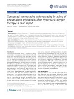
báo cáo khoa học: "Computed tomography colonography imaging of pneumatosis intestinalis after hyperbaric oxygen therapy: a case report" docx
Ngày tải lên :
10/08/2014, 22:24
... view of surface rendering computed tomography colonography (CTC) image demonstrating multiple gas cysts in the left colon and sigmoid (B) Fly-through view of CTC of the sigmoid (C) Coronal view of ... treating PI and has the major advantage of avoiding the pulmonary toxicity of oxygen that can be associated with use of prolonged high flow oxygen [9] The use of heliox seems to be more effective ... this article as: Frossard et al.: Computed tomography colonography imaging of pneumatosis intestinalis after hyperbaric oxygen therapy: a case report Journal of Medical Case Reports 2011 5:375...
- 4
- 340
- 0
báo cáo khoa học: "Successful treatment of a free-moving abdominal mass with radiation therapy guided by conebeam computed tomography: a case report" potx
Ngày tải lên :
11/08/2014, 02:21
... cone-beam computed tomography had not been used
Dabaja et al Journal of Medical Case Reports 2010, 4:329 http://www.jmedicalcasereports.com/content/4/1/329 Page of Abbreviations CT: computed tomography; ... 77030, USA 3Department of Radiation Physics, The University of Texas MD Anderson Cancer Center, Houston, Texas 77030, USA Figure Positron emission tomography /computed tomography images obtained ... al Journal of Medical Case Reports 2010, 4:329 http://www.jmedicalcasereports.com/content/4/1/329 Page of with conventionally planned treatment ports because of the extensive motion of the tumor...
- 4
- 375
- 0

báo cáo khoa học: " Computed tomography imaging of subpleural lipoma in two men: two case reports" potx
Ngày tải lên :
11/08/2014, 02:22
... Journal of Medical Case Reports 2010, 4:380 http://www.jmedicalcasereports.com/content/4/1/380 Page of Figure Axial computed tomography (CT) image showing the characteristic hypodense condition of ... lipomas Figure Axial computed tomography (CT) image in mediastinal soft tissue window-level setting shows the characteristic appearance of a fat-containing tumor with density values of approximately ... reported before the examination were caused by recurrent attacks of bronchitis Discussion Before the implementation of computed tomography in diagnostic medical imaging, most subpleural lipomas...
- 4
- 279
- 0

Báo cáo y học: "Role of positron emission tomography-computed tomography in bronchial mucoepidermoid carcinomas: a case series and review of the literature" pdf
Ngày tải lên :
11/08/2014, 03:21
... bronchial mucoepidermoid carcinoma Axial section of positron emission tomography (PET), computed tomography and PET -computed tomography images of 18F FDG scan showing mild FDG uptake in the histologically ... bronchial mucoepidermoid carcinoma Axial section of positron emission tomography (PET), computed tomography and PET -computed tomography images of 68Ga DOTATOC scan showing no significant radiotracer ... Phel1-Tyr3-octreotide positron emission tomography computed tomography) , F- Female, FDG PET-CT- fluorodeoxyglucose positron emission tomography computed tomography, H-Haemoptysis, M- Male, MECmucoepidermoid...
- 4
- 210
- 0

Báo cáo y học: "Soft-tissue perineurioma of the retroperitoneum in a 63-year-old man, computed tomography and magnetic resonance imaging findings: a case report" doc
Ngày tải lên :
11/08/2014, 03:21
... Yasumoto et al Journal of Medical Case Reports 2010, 4:290 http://www.jmedicalcasereports.com/content/4/1/290 Page of Figure Computed tomography results (a) Plain computed tomography (CT) shows ... The immunohistochemical studies are often necessary for the diagnosis of soft-tissue perineurioma [3,4] To the best of our knowledge, MRI and CT images of soft-tissue perineurioma in the retroperitoneum ... one of the MR imaging characteristics of ganglioneuroma is curvilinear bands of low signal intensity on T2-weighted images [7], which were absent in the current soft-tissue perineurioma The soft-tissue...
- 4
- 384
- 0
Tìm thêm:
- hệ việt nam nhật bản và sức hấp dẫn của tiếng nhật tại việt nam
- xác định các mục tiêu của chương trình
- xác định các nguyên tắc biên soạn
- khảo sát các chuẩn giảng dạy tiếng nhật từ góc độ lí thuyết và thực tiễn
- khảo sát chương trình đào tạo của các đơn vị đào tạo tại nhật bản
- khảo sát chương trình đào tạo gắn với các giáo trình cụ thể
- xác định thời lượng học về mặt lí thuyết và thực tế
- tiến hành xây dựng chương trình đào tạo dành cho đối tượng không chuyên ngữ tại việt nam
- điều tra đối với đối tượng giảng viên và đối tượng quản lí
- điều tra với đối tượng sinh viên học tiếng nhật không chuyên ngữ1
- khảo sát thực tế giảng dạy tiếng nhật không chuyên ngữ tại việt nam
- khảo sát các chương trình đào tạo theo những bộ giáo trình tiêu biểu
- nội dung cụ thể cho từng kĩ năng ở từng cấp độ
- xác định mức độ đáp ứng về văn hoá và chuyên môn trong ct
- phát huy những thành tựu công nghệ mới nhất được áp dụng vào công tác dạy và học ngoại ngữ
- mở máy động cơ lồng sóc
- mở máy động cơ rôto dây quấn
- các đặc tính của động cơ điện không đồng bộ
- hệ số công suất cosp fi p2
- đặc tuyến hiệu suất h fi p2