basic structure of the quran


Tài liệu Báo cáo khoa học: Crystal structure of the cambialistic superoxide dismutase from Aeropyrum pernix K1 – insights into the enzyme mechanism and stability pdf
... the ter- tiary structure of ApeSOD has not been elucidated. In the present study, for the first time, we describe the crystal structure of ApeSOD. In particular, we focus on the coordination of ... ligand, NE2 of His31, bound to the metal, in the company of a water oxygen, from the apical positions. The manga- nese was only 0.06 A ˚ out of the equatorial plane (Table 3). The angles around the metal ... metal cofactor. Figure 3D shows the superimposition of the active site structures of apo, Mn-bound and Fe-bound Ape- SODs. The most significant difference among them was related to the OH of Tyr39,...
Ngày tải lên: 14/02/2014, 22:20

Tài liệu Inequalities in Higher Education and the Structure of the Labour Market pdf
... especially thosewhoholdgeneralbachelor’sdegrees,theirqualificationsserveonlyasasignal of theirpotentialtoemployers.Inmanycases,theytakelongertorealiseemployment andwhentheydotheirentryleveljobsdonotnecessarilyrequire the years of schooling theypossess.Theystartat the bottom of the jobladderandhavetoprovethemselves in the labourmarkettoreachhigherincomelevels.Acombination of theirpotential andtrainingwilldeterminetheirprogressionup the ladder.Aswillbeseenin the next section, the field of study,whichsignals the type of skillsacquiredthrougheducation, playsasignificantroleinemployability of universitygraduatesinthiscase. PercyMoleke Free ... www.hsrcpress.ac.za 4 PercyMoleke 5 InequalitiesinHigherEducationand the Structure of the LabourMarket Despite the observedsignificantincreasesin the number of seniorcertificateholders in the pastdecade, the proportion of learnerswhoactuallysitfor the examremains low. The quality of the seniorcertificateexamalsocontinuestobe of concern. The largemajority of learnersstillopttowrite the seniorcertificateexamon the standard graderatherthanon the highergrade.Consequently, the proportion of thosepassing with ... www.hsrcpress.ac.za 14 PercyMoleke 15 InequalitiesinHigherEducationand the Structure of the LabourMarket ineducationorin the labourmarket,havedonelittletoaddress the inequalities of the past. The lack of labour marketinformation...
Ngày tải lên: 15/02/2014, 17:20
Tài liệu High-level Expert Group on reforming the structure of the EU banking sector docx
... to the intensification of the sovereign debt crisis in the euro area. The pricing of risk in the repo market has become more dependent on the geographic origin of both the counterparty and the ... maintaining the integrity of the single market. Finally, Chapter 5 draws together the analysis of the previous chapters. It reiterates the importance of banks in the EU economy, summarises the key ... mandate, the Group also expects the on-going fundamental review of the trading book by the Basel Committee to improve the control of market risk within the banking system. The Group sees the Commission's...
Ngày tải lên: 16/02/2014, 10:20

Tài liệu Báo cáo khoa học: Structure of the putative 32 kDa myrosinase-binding protein from Arabidopsis (At3g16450.1) determined by SAIL-NMR docx
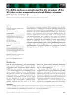
... of SAIL Fig. 3. Secondary structure of At3g16450.1. (A) Ribbon representa- tion of the NMR structure of At3g16450.1. These figures were pre- pared using MOLMOL [25]. Due to the lack of NOEs, the ... constraints. Solution structure of SAIL At3g16450.1 Assignment of the NOE peaks of At3g16450.1 and the structure determination were accomplished by use of the cyana program [17,18]. The structural statistics ... Three-dimensional NMR structure of At3g16450.1. (A) Superposition of the 20 energy-minimized conformers that repre- sent the 3D solution structure of the N-terminal domain. (B) Super- position of conformers...
Ngày tải lên: 18/02/2014, 14:20

Tài liệu Báo cáo khoa học: Crystal structure of the catalytic domain of DESC1, a new member of the type II transmembrane serine proteinase family pptx
... Asp189 at the bottom of the pocket determines the specificity of the S1 pocket for basic residues Arg and Lys at position P1 of the substrate. Consequently, in the DESC1 complex structure the bound ... reveals that the most similar regions of these proteinases medi- ate interaction of the two b-barrels, formation of the catalytic machinery and structures required for binding of the main chain of the ... below). The southern boundary of the active site cleft of DESC1 is formed by the 145 autolysis loop. The backbone of this loop differs mark- edly from the other serine proteinases, making the act- ive...
Ngày tải lên: 19/02/2014, 00:20

Tài liệu Báo cáo khoa học: Crystal structure of the BcZBP, a zinc-binding protein from Bacillus cereus doc
... determines the shape of the active site entry. (e) The structure of the active site is essentially identical with the active sites of the MshB and LpxC proteins. The conservation of catalytically important ... studying the corresponding proteins of B. anthracis because it lacks infectiousness of the latter. The objective of the present study is to shed light on the structure, function and the structure function ... diagram. B A Fig. 1. Structure of the BcZBP dimer. (A) Topology diagram of the dimer drawn with the program TOPDRAW [34]. The relative orienta- tion of the secondary structure elements is illustrated. The shaded areas...
Ngày tải lên: 19/02/2014, 00:20

Tài liệu Báo cáo khoa học: Spectroscopic characterization of a higher plant heme oxygenase isoform-1 from Glycine max (soybean) ) coordination structure of the heme complex and catabolism of heme docx
... information on the heme pocket structure. As shown in Table 2, the hyperfine splitting constants of the 15 N nucleus of the distal NO and of the 14 N nucleus of the proximal His of the GmHO-1 complex ... against the concentration of azide of each solution. The intersection point corresponding to the mole fraction of 0.5 of the respective forms gives the amount of azide necessary to attain the equilibrium, ... characteristics of the heme pocket of GmHO-1 might be associated with the smaller values of K azide for GmHO-1 and SynHO-1, and with the ferric character of the heme. The K imidazole values of the three...
Ngày tải lên: 19/02/2014, 05:20

Tài liệu Báo cáo khoa học: Unusual metal specificity and structure of the group I ribozyme fromChlamydomonas reinhardtii23S rRNA pptx
... the large P6 extension and the last three nucleotides (AU XG) of the intron. The sizes of the intron (InDG b ) and 5¢ exon (5E) are indicated. The arrow indicates cleavage of the pre-RNA at the ... Because of these and other properties of Cr.LSU, the tertiary structure of the intron from 23S.5DG b was examined using Fe 2+ -EDTA cleavage. The ground-state structure shows evidence of an unusually ... 23S.5DG b pre-RNA, and the binding of a minimum of n Mg 2+ ions leads to formation of the active complex, whereas binding of a minimum of m Mn 2+ ions forms the inactive complex. The sizes of the active and inactive...
Ngày tải lên: 19/02/2014, 07:20

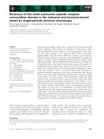
Tài liệu Báo cáo khoa học: Proton transfer in the oxidative half-reaction of pentaerythritol tetranitrate reductase Structure of the reduced enzyme-progesterone complex and the roles of residues Tyr186, His181 and His184 pdf
... confirming that the mutation of Tyr186 to Phe186 does not grossly perturb the overall framework of the enzyme or the active site. The active sites of the enzymes are shown in stick format with the sigmaA ... productive binding of steroid substrates: in the first, a (small) sub population of enzyme binds directly the steroid in the reactive conformation, with the remainder of the enzyme binding the unreactive conformation. ... bond in close proximity of the N5 atom. A putative flipping motion of the progesterone molecule along an axis parallel to the FMN plane aligns the C1-C2 with the plane of the isoalloxazine ring...
Ngày tải lên: 20/02/2014, 02:21
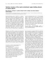
Tài liệu Báo cáo khoa học: Solution structure of the matrix attachment region-binding domain of chicken MeCP2 ppt
... [18]. The structure of the helical coil a 2 /a 3 allows us to interpret the consequences of the six mutations. As P153(152) and G162(161) are buried in the protein core, replacement of each of these ... one-turn helix on the other face. It is thought that the two inner strands of the b-sheet lie within the major groove of the DNA and that a hydrophobic pocket formed by the side chains of Y123 and ... subsequent structure calculations. These newly calculated structures were then used for the next step in the iteration process. This procedure was continued until the quality of the structure could...
Ngày tải lên: 21/02/2014, 00:20

Tài liệu Báo cáo khoa học: The structure of the carbohydrate backbone of the lipopolysaccharide from Acinetobacter baumannii strain ATCC 19606 docx
... 269) 427 [26,27]. Then, the biosynthesis of the core region of LPS from A. baumannii strain ATCC 19606 should proceed with the introduction of a third Kdo residue to O-5 of the second Kdo. This ... [19]. The mechanism of the transfer of the third Kdo residue has not yet been identi®ed. In addition to the work on the determination of the chemical and antigenic structures of O-speci®c polysaccha- rides ... genus which obviously allows novel insights in structure, genetics and biosynthesis of LPS. Here, the structures of the carbohy- drate backbones of the LPS from A. baumannii strain ATCC 19606 are...
Ngày tải lên: 21/02/2014, 03:20

Tài liệu Báo cáo Y học: Structure of the O-polysaccharide and classification of Proteus mirabilis strain G1 in Proteus serogroup O3 potx
... has the D configuration. On the basis of the data obtained, it was concluded that the O-polysaccharide P. mirabilis G1 has the structure shown in Fig. 3. This structure is similar to that of the O-polysaccharide ... experi- ments. The following structure of the polysaccharide was established: where D -GalA6( L -Lys) stands for N a -( D -galacturonoyl)- L -lysine. The structure of the O-polysaccharide of P. mirabilis ... relatedness of P. mirabilis G1 and S 1959 correlated with the similarity of the chemical structures of their O-polysaccharides (Fig. 3). Therefore, it is reasonable to classify P. mirabilis G1 into the...
Ngày tải lên: 21/02/2014, 15:20

Tài liệu Báo cáo Y học: Solution structure of the mEGF/TGFa44250 chimeric growth factor doc
... found. Several of these were identified in previous studies of EGF, and the backbone fold of the chimera is clearly similar to those of the EGF structures. The presence of a backbone hydrogen bond from the ... structure from each of the previously reported families of structures for mEGF; the results are given in Table 3. The difference between the backbone of the chimera and the EGF structure is no greater ... the calculation of the chimera structure, this provides strong evidence that the overall structure is unchanged by the modification of the sequence. Given that the solution structure of mEGF/TGFa 44250 was...
Ngày tải lên: 22/02/2014, 07:20

Báo cáo khoa học: Solution structure of the catalytic domain of RICH protein from goldfish pot
Ngày tải lên: 07/03/2014, 10:20
Bạn có muốn tìm thêm với từ khóa: