PARKINSON’S DISEASE AND RELATED DISORDERS - PART 6 ppt

PARKINSON’S DISEASE AND RELATED DISORDERS - PART 6 ppt
... striato- nigral degeneration showing sym- metrical atrophy and discoloration of the putamen Figure 32 11 C-raclopride binding in a normal subject (left) compared with that in Parkinson's disease (middle) ... atrophy ©2004 CRC Press LLC Figure 42 T 2 -weighted MRI (upper) shows hyperintensity of the middle cere- bellar peduncles and the cerebellum. The axial proton-density MRI (low...
Ngày tải lên: 09/08/2014, 16:21
PARKINSON’S DISEASE AND RELATED DISORDERS - PART 8 ppt
... LLC Figure 66 T 1 -weighted MRI shows the presence of high-signal areas (arrowed) in the pallidum in a patient with chronic acquired hepatocere- bral degeneration ©2004 CRC Press LLC Figure 64 Histology ... PETscans showing integrated 11 C-raclopride and 11 C-SCH 23390 activity in a normal subject (left) and choreic patient (right) with Huntington's disease. Both D 1 and D 2 b...
Ngày tải lên: 09/08/2014, 16:21

PARKINSON’S DISEASE AND RELATED DISORDERS - PART 5 pot
... normal) arcuate zone (white arrow), and rarefied pale-staining deep white matter, containing thick-walled arteriosclerotic blood vessels lying in dilated and fibrotic perivascular spaces (black ... 15 6- [ 18 F]-fluorodopa–PET scan appearance in a normal subject (upper) compared with a Parkinsonian patient (lower) Figure 14 Positive glabellar tap. Persistent blinking is a feature of Pa...
Ngày tải lên: 09/08/2014, 16:21
PARKINSON’S DISEASE AND RELATED DISORDERS - PART 7 pot
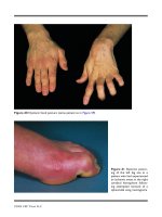
... Dorsal (left) and palmar (right) views of dystonic posturing of the left hand of a patient with corticobasal degeneration. In particular, note the ulnar deviation at the wrist and the abducted ... 57 Dystonic postur- ing of the hand consequent to perinatal hypoxia Figure 58 Dystonic postur- ing of the foot consequent to perinatal hypoxia. There is inversion of the foot and relative dors...
Ngày tải lên: 09/08/2014, 16:21

Tài liệu Parkinson’s Disease and Related Disorders docx
... 43 Methods Subjects Seven right-handed L-Dopa-treated patients with clini- cally definite mild-moderate PD (Hoehn & Yahr stage 1–2 (Hoehn and Yahr, 1 967 )) were recruited, and were asked to use their right hand. All ... in the alpha- and beta frequency bands. For instance, in parkinso- nian monkeys and patients, oscillatory burst- ing typically emerges in both the 3–8 Hz band, an...
Ngày tải lên: 14/02/2014, 17:20