Parkinson disease and related disorders part - part 1 ppsx

PARKINSON’S DISEASE AND RELATED DISORDERS - PART 5 pot
... LLC Figure 15 6-[ 18 F]-fluorodopa–PET scan appearance in a normal subject (upper) compared with a Parkinsonian patient (lower) Figure 14 Positive glabellar tap. Persistent blinking is a feature of Parkinson s ... left hand Figure 13 Power-spectrum (upper) and accelerometer (lower) tracings taken from a patient with Parkinsonian tremor. The main tremor peak is at approximately 5 Hz...
Ngày tải lên: 09/08/2014, 16:21

PARKINSON’S DISEASE AND RELATED DISORDERS - PART 6 ppt
... striato- nigral degeneration showing sym- metrical atrophy and discoloration of the putamen Figure 32 11 C-raclopride binding in a normal subject (left) compared with that in Parkinson& apos;s disease (middle) ... with the doll's-head maneuver (right) in this patient with progressive supranuclear palsy Figure 31 Sagittal T 1 -weighted MRI showing midbrain atrophy (arrowed) in...
Ngày tải lên: 09/08/2014, 16:21
PARKINSON’S DISEASE AND RELATED DISORDERS - PART 7 pot
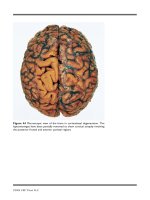
... The leptomeninges have been partially removed to show cortical atrophy involving the posterior frontal and anterior parietal regions ©2004 CRC Press LLC Figure 51 [ 18 F]-fluorodeoxyglucose– PET ... extension and dilatation of the right frontal horn (white arrow) ©2004 CRC Press LLC Figure 50 In this patient with corticobasal degenera- tion, sagittal T 1 -weighted MRI (upper) shows p...
Ngày tải lên: 09/08/2014, 16:21
PARKINSON’S DISEASE AND RELATED DISORDERS - PART 8 ppt
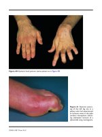
... showing integrated 11 C-raclopride and 11 C-SCH 23390 activity in a normal subject (left) and choreic patient (right) with Huntington's disease. Both D 1 and D 2 binding is reduced in the ... (arrow- ed). The coronal proton-density MRI (lower) shows a similar picture together with an anteromedial zone of high intensity ©2004 CRC Press LLC Figure 73 PETscans showing integrate...
Ngày tải lên: 09/08/2014, 16:21
Tài liệu Parkinson’s Disease and Related Disorders docx
... 205 Dawson, T. M.: Parkin and defective ubiquitination in Parkinson s disease 209 Heutink, P.: PINK -1 and DJ -1 – new genes for autosomal recessive Parkinson s disease . . . 215 Whaley, N. R., Uitti, ... among primary motor cortex neurons in the 1- methyl- 4- phenyl -1 , 2,3,6-tetrahydropyridine primate model of Parkinson s disease. J Neurosci 22: 4639 –4653 Guzman JN, Her...
Ngày tải lên: 14/02/2014, 17:20