classification of diabetic foot infections

2012 Infectious Diseases Society of AmericaClinical Practice Guideline for the Diagnosisand Treatment of Diabetic Foot Infections
... patients with diabetic foot infections Foot Ankle Int 1998; 19: 38–40 Mueller-Buehl U, Diehm C, Gutzler F, Adam D Tissue concentrations of ofloxacin in necrotic foot lesions of diabetic and non -diabetic ... deeper insight into diabetic foot care? J Foot Ankle Surg 2006; 45:375–9 270 Jones V Debridement of diabetic foot lesions The Diabetic Foot 1998; 1:88–94 271 Rauwerda JA Foot debridement: anatomic ... management of diabetes-related foot ulceration: a pilot study Adv Skin Wound Care 2004; 17:232–8 Aragon-Sanchez J Seminar review: a review of the basis of surgical treatment of diabetic foot infections...
Ngày tải lên: 14/04/2016, 19:08

Báo cáo y học: "Classification of hip joint infections"
... joint [8] This classification system consists of stages (Table 1) and combines intraarticular findings of the soft tissues as well as radiological alterations of the infected joint Infections classified ... requires open revision surgery Table 1: Arthroscopic classification of joint infections according to Gächter [8] Stage I opacity of fluid, redness of the synovial membrane, possible petechial bleeding, ... the Cierny classification system for osteomyelitis in adult patients [4] also for the classification of periprosthetic total joint infections [3] In this system, prosthetic joint infections are...
Ngày tải lên: 26/10/2012, 09:57

The clinical study and the prevention countermeasures of diabetic foot
... Preventing complications of diabetes, especially improving awareness of Diabetic foot, studying methods of prevention and treatment are very important Key words: Diabetic foot, clinical study , ... modality of treatment for diabetic foot in Institute of Traditional medicine in Hochiminh City Method: Aspect of basic theory: Collecting and summarizing Western and Chinese medicine documents in diabetic ... pattern identification of diabetic foot, making progress to perfect clinical syndrome differentiation and treatment for diabetic foot, thenceforward dignifying knowledge of traditional Chinese...
Ngày tải lên: 22/05/2016, 16:56

Atlas of the Diabetic Foot - part 1 pot
... Atlas of the Diabetic Foot Professor Nicholas Katsilambros, MD Director of the 1st Department of Propaedeutic Medicine and the Diabetic Centre Athens University Medical ... Chapter VIII Infections Chapter IX Neuro-Osteoarthropathy The Charcot Foot vii ix 23 41 73 85 105 125 151 185 Appendix Anatomy of the Foot Appendix Manufacturers of Preventive and Therapeutic Footwear ... specialists in infectious diseases or orthopedics or as scholars in the field of diabetes and the diabetic foot Atlas of the Diabetic Foot N Katsilambros, E Dounis, P Tsapogas and N Tentolouris Copyright...
Ngày tải lên: 10/08/2014, 18:21

Atlas of the Diabetic Foot - part 2 ppsx
... PATIENTS IN APPROPRIATE FOOT CARE Education of patients who are at risk of developing foot ulceration is the cornerstone of disease management Patients 32 Atlas of the Diabetic Foot should fully understand ... treatment of neuropathic ulcers with promising results Keywords: Classification of foot ulcers; Meggitt–Wagner classification of foot ulcers; ‘The University of Texas classification system for diabetic foot ... The Diabetic Foot (6th edn) St Louis: Mosby, 2001; 273–282 Macfarlane RF, Jeffcoate WJ Classification of foot ulcers: The S(SAD)SAD system Diabetic Foot 1999; 2: 123–131 40 Atlas of the Diabetic...
Ngày tải lên: 10/08/2014, 18:21

Atlas of the Diabetic Foot - part 3 pdf
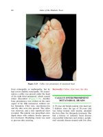
... Atlas of the Diabetic Foot Figure 3.31 Photograph of the foot shown in Figures 3.28–3.30 months after arthrodesis of the first metatarsophalangeal joint and second ray amputation Note the absence of ... the arch of the foot is higher than normal (pes cavus) claw toes often develop In cavus foot the forefoot, and especially the first ray, is drawn downwards and an abnormal distribution of plantar ... and hemorrhagic calluses of the second and third toes were also present A hammer deformity was seen on the second toe of her right foot Protective 48 Atlas of the Diabetic Foot Figure 3.8 Curly...
Ngày tải lên: 10/08/2014, 18:21
Atlas of the Diabetic Foot - part 4 doc
... the lateral aspect of the foot of a diabetic patient The design of the excision and the recipient vessels are indicated (Courtesy of O Papadopoulos) 82 Atlas of the Diabetic Foot Figure 4.14 Free ... excision of a dermatofibrosarcoma protuberans of heel Patient of Figure 4.15 (Courtesy of O Papadopoulos) Figure 4.15 Ulceration of recurrent dermatofibrosarcoma protuberans of the heel of a diabetic ... (on the right foot at the age of 66 years and on the left foot at the age of 68 years) because Anatomical Risk Factors for Diabetic Foot Ulceration of infected foot ulcers under the metatarsal...
Ngày tải lên: 10/08/2014, 18:21

Atlas of the Diabetic Foot - part 5 potx
... Atlas of the Diabetic Foot Figure 5.23 Plain radiograph of the right foot of the patient whose foot is shown in Figure 5.21 Osteomyelitis of the fifth metatarsal head and the proximal phalanx of ... 5.19 Hindfoot shown in Figure 5.17 after the ulcer has completely healed Figure 5.18 Commercially available heel-free shoes for the treatment of hindfoot ulcers 98 Atlas of the Diabetic Foot Figure ... ULCERS OVER A CHARCOT FOOT The following two figures (before and after debridement) show the left foot of a male patient of 62 years of age with type diabetes diagnosed at the age of 48 years and treated...
Ngày tải lên: 10/08/2014, 18:21

Atlas of the Diabetic Foot - part 6 ppsx
... whose foot is shown in Figure 6.14 The yellowish appearance of the bed of the ulcer is indicative of ischemia 116 Atlas of the Diabetic Foot of 55 years, was referred to the outpatient diabetic foot ... age of 42 years and being treated with sulfonylureas, was referred to the outpatient diabetic foot clinic 134 Atlas of the Diabetic Foot Figure 7.9 Digital subtraction angiography of the foot ... gangrene of the distal areas of the toes of the right foot The well-demarcated red area extending up to the ankle and the lateral foot indicates ischemic necrosis of the skin (Courtesy of E Bastounis)...
Ngày tải lên: 10/08/2014, 18:21

Atlas of the Diabetic Foot - part 7 ppt
... Atlas of the Diabetic Foot pathogens that colonize the skin of diabetic patients, are the causative agents of acute infections in antibiotic-na¨ve patients, ı and are nearly always the cause of ... gangrene of the right foot Redness and edema due to infection extends as far as the lower third of the tibia (Courtesy of E Bastounis) Gangrene 145 Figure 7.24 Wet gangrene of midfoot and forefoot ... complained of pain in his right foot when he was at rest, weeks prior to 144 Atlas of the Diabetic Foot Figure 7.21 Wet gangrene of the last two toes An infected area of ischemic necrosis is also...
Ngày tải lên: 10/08/2014, 18:21

Atlas of the Diabetic Foot - part 8 ppt
... and severe neuropathy 162 Atlas of the Diabetic Foot Figure 8.10 Deep tissue infection of the foot following web space mycosis Redness and edema of the whole foot with pustules on the dorsum ... patient with type diabetes mellitus since the age of 40 years attended the diabetic foot clinic because of a large ulcer of the sole of her left foot She was being treated with insulin resulting ... ulcer after month of chiropody treatment Healthy granulated tissue covers the bed of the ulcer 166 Atlas of the Diabetic Foot Figure 8.16 Left neuro-osteoarthropathic foot of the patient whose...
Ngày tải lên: 10/08/2014, 18:21

Atlas of the Diabetic Foot - part 9 pptx
... resorption of phalanges and 190 Atlas of the Diabetic Foot Figure 9.2 Radiograph of acute neuro-osteoarthropathy showing osteolytic destruction of the third and fourth metatarsal heads, widening of the ... collapse of the midfoot A minimal (of a few millimeters) lateral deviation or a fracture of the base of the second metatarsal may be an early sign of acute neuro-osteoarthropathy If the foot is ... of 46 years and acceptable diabetes control (HBA1c : 7.4%), was referred to the outpatient diabetic foot clinic for a chronic plantar ulcer on his left midfoot The 202 Atlas of the Diabetic Foot...
Ngày tải lên: 10/08/2014, 18:21

Atlas of the Diabetic Foot - part 10 pps
... ANATOMY OF THE FOOT Anatomy of the Foot 215 Figure A1 Dorsal aspect of the bones in the foot Figure A2 the foot Figure A3 Plain radiograph of the foot shown in lateral view Plantar aspects of the ... 208, 209 flat foot see pes planus Fontaine clinical staging 7–8 neuro-ischemic ulcers 116 224 Index foot anatomy 215 see also forefoot; heel; hindfoot; midfoot; sole of foot; toe(s) foot care, patient ... Lateral view of Figure 9.29 Figure 9.31 Plain radiograph of neuro-osteoarthropathy of the right foot of the patient whose feet are shown in Figures 9.29 and 9.30 There is complete destruction of the...
Ngày tải lên: 10/08/2014, 18:21

Báo cáo y học: "Diagnostic accuracy of existing methods for identifying diabetic foot ulcers from inpatient and outpatient datasets" pptx
... Sohn et al Journal of Foot and Ankle Research 2010, 3:27 http://www.jfootankleres.com/content/3/1/27 Page of Table Comparison of methods for diagnostic accuracy of diabetic foot ulcers (N = 518) ... specificity Sohn et al Journal of Foot and Ankle Research 2010, 3:27 http://www.jfootankleres.com/content/3/1/27 Page of Table Existing methods of identifying diabetic foot ulcers in administrative ... According to this method, the annual prevalence of diabetic foot ulcers was 3.3% in the VA diabetic population in 2003 List of abbreviations DFU: Diabetic foot ulcers; NPV: negative predictive value;...
Ngày tải lên: 10/08/2014, 21:24
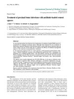
Báo cáo y học: "Treatment of proximal femur infections with antibiotic-loaded cement spacers"
... provision of sufficient infection therapy is the usage of temporary, antibiotic-loaded cement spacers [5, 7, 14, 24] Although their indication in the treatment of destructive, bacterial infections of ... Fig 2: Evaluation of the hip joint function by the Merle d’ Aubigne score at the site of spacer implantation in the treatment of proximal femur infections Fig 3: Evaluation of the hip joint function ... for an adequate option in the treatment of periprosthetic infections Although their indication for the treatment of destructive, bacterial infections of the proximal femur would make sense, literature...
Ngày tải lên: 26/10/2012, 09:53

Báo cáo y học: "Complications after spacer implantation in the treatment of hip joint infections"
... properties of gentamicin-loaded hip spacers after insertion of Kirschner wires [31] Stress experiments showed an average failure load of 1.6 kN The insertion of the K-wires prevented any dislocation of ... a study of 274 patients who underwent total hip replacement that of the patients with deep joint sepsis who had preoperative urinary tract infections, had evidence of the same organism of both ... retrospective evaluation of the patients’ records did not allow any differentiation of the particular cause of the renal failure, respectively (antibiotic-impregnation of the spacer, systemic...
Ngày tải lên: 26/10/2012, 09:53

Báo cáo y học: "Revision of late periprosthetic infections of total hip endoprostheses: pros and cons of different concepts"
... small numbers in the form of a biofilm and are also often in a sessile state that is characterized by a slow rate of reproduction [8,10-13] An analysis we carried out of 110 infected hip and knee ... lack of sufficient incubation led to the poor sensitivity of the pre-operative aspiration reported in other studies (for example, 46.1% reported by Hoffmann et al [17]) The degree of success of ... cement of the spacer is not intended as a means of fixing the prosthesis so the mechanical characteristics of the cement is not of primary importance at this stage Thus, large amounts of antibiotics...
Ngày tải lên: 26/10/2012, 09:53

iec 60079-10 electrical apparatus for explosive gas atmospheres - classification of hazardous areas
... adjacent sources of release overlap and are of different zonal classification, the higher risk classification will apply in the area of overlap Where overlapping zones are of the same classification, ... Type of zone The likelihood of the presence of an explosive gas atmosphere and hence the type of zone depends mainly on the grade of release and the ventilation NOTE A continuous grade of release ... pressure and the geometry of the source of release The size of a cloud of flammable gas or vapour is determined by the rate of flammable vapour release and the rate of dispersion Gas and vapour...
Ngày tải lên: 25/12/2013, 10:41
Tài liệu Báo cáo " DETERMINING FLOW ENERGY AND EROSION COEFFICIENT FOR CLASSIFICATION OF POTENTIAL EROSION IN BINH DINH PROVINCE " ppt
... CSIC, 1998) The results of erosion potential classification - Based on Binh Dinh profile’s file (Vietnam Association of Soil Sciences, 1997), values of K for 117 hill soil profiles throughout the ... coefficient 39 The method of erosion potential classification in Binh Dinh Province The classification of erosion potential M in Binh Dinh Province is based on the numerical value of the flow energy ... of erosion potential for all pixels of the four basins Table K values of main soil groups and classification by erosive characteristics Symbology Number of profiles Pc-h Speck aluvial soil Pc-fe...
Ngày tải lên: 13/02/2014, 12:20

Tài liệu Gait Pattern Classification of Healthy Elderly Men Based on Biomechanical Data ppt
... developed of the Yl family in the (A) sagittal, is overlaid in bold distance of 8.7 and 13.7, respectively They form one family of 10 trials, El, and a smaller one, E2, of 6, all consisting of elderly ... center of a 10-m walkway to cover the volume required for at least one complete gait cycle of the right lower limb A third camera was placed in front of the subject and aligned along the axis of ... side), in terms of the effect of the combined 31 gait parameters The optimal number of clusters can be determined in several ways Winters and coworkers*’ calculated the criterion of Hartigan and...
Ngày tải lên: 14/02/2014, 07:20