applications of atomic force microscopy in nanomaterials research

Atomic Force Microscopy in Cell Biology_Methods in Cell Biology Volume 68 pptx
... understanding of how nanomachines, like kinesin, convert chemical energy into mechanical movement. So far, attempts to satisfactorily explain the function of kinesin using the knowledge gained by ... and kinematics in addition to the application of other measuring techniques possible. With this step in the development of scanning probe instruments, the capability of optical microscopy to investigate ... specific binding, for instance, to membrane and other molecular structures for a certain time and to track their motion. A. Mechanics of Molecular Motors Kinesins and kinesin-like proteins are of wide...
Ngày tải lên: 27/06/2014, 08:20

Atomic Force Microscopy in Cell Biology ppt
... understanding of how nanomachines, like kinesin, convert chemical energy into mechanical movement. So far, attempts to satisfactorily explain the function of kinesin using the knowledge gained by ... for instance, to membrane and other molecular structures for a certain time and to track their motion. A. Mechanics of Molecular Motors Kinesins and kinesin-like proteins are of wide interest in ... structure that interacts with its light chain, binding to the cargo. Several studies on kinesins have shown that the neck and the first hinge region of the motor play important roles in kinesin directionality,...
Ngày tải lên: 27/06/2014, 17:20

Atomic Force Microscopy in Cell Biology Episode 1 Part 1 ppt
... Ricci Atomic Force Microscopy Biomedical Methods and Applications Imaging Methods in AFM 21 Form the point of view of biomedical applications, interesting experiments can be performed by coating ... number of data points per line, obtain- ing at the end a square grid of data points each corresponding to the relative x, y, and z coordinates in space of the sample surface (11). Usually during ... Imaging Methods in AFM 13 13 2 Imaging Methods in Atomic Force Microscopy Davide Ricci and Pier Carlo Braga 1. Introduction One can easily distinguish between two general modes of operation of...
Ngày tải lên: 06/08/2014, 02:20
Atomic Force Microscopy in Cell Biology Episode 1 Part 2 pptx
... test pattern (as in Fig. 4) will produce an asymmetric profile. In the case of contaminants, one often notices an abrupt change of detail contrast during scanning and a blurring of the image. Sometimes ... atomic force microscopy tips be inspected by atomic force microscopy? J. Vac. Sci. Technol. B. 9, 1309–1312. 3. Keller, D. and Chou, C. C. (1992) Imaging steep, high structures by scanning force microscopy ... by inducing stress on one surface of the cantilever as the result of denser packing of the molecules (8) or a frequency shift in case of dynamic detection mode (9) as a result of changes in mass. 3....
Ngày tải lên: 06/08/2014, 02:20

Atomic Force Microscopy in Cell Biology Episode 1 Part 3 pdf
... washed again in PBS before fixing in 0.01% gluteraldehyde (in S buffer) for 10 min. This is followed by another wash in PBS before being thoroughly rinsed in deionized and distilled water. Finally, ... As in the cases described in Subheading 3.1.2., a soft lever (k N < 0.01 N/m) in combination with a low applied force (<1 nN) will enhance information arising from the softer elements of ... strategy to gain insight into cytoskeletal dynamics (e.g., refs. 5,12–14). An example of investigations of slow intracellular dynamics is shown in a sequence of images in Fig. 3, where the intracellular...
Ngày tải lên: 06/08/2014, 02:20
Atomic Force Microscopy in Cell Biology Episode 1 Part 4 potx
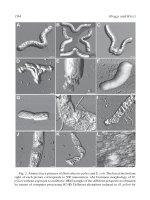
... imaging difficulties. The inter- action of glycoaminoglycans chains of the glycocalyx layer with the microscope tip makes the imaging of the fresh tissue difficult (Fig. 6). When the imaging of a ... unevenness of the sample sur- face thus increasing contrast. In addition, information concerning the sample’s molecular composition can be revealed and help in the interpretation of the effect of the ... cells measured by atomic force microscopy: effects of anticytoskeletal drugs and mem- brane crosslinking. Scanning 20, 389–397. 20. Kuznetsov, Y. G., Malkin, A. J., and McPherson, A. (1997) Atomic force...
Ngày tải lên: 06/08/2014, 02:20

Atomic Force Microscopy in Cell Biology Episode 1 Part 5 doc
... 95 95 8 Calculation of Cuticle Step Heights from AFM Images of Outer Surfaces of Human Hair James R. Smith 1. Introduction Atomic force microscopy (AFM) is an ideal technique for noninvasive examination of hair ... providing a wealth of structural informa- tion not always apparent from electron microscopy. The fine cuticular struc- ture of human head hair is of interest to those engaged in the fields of dermatology ... first line consisting of 500 data points of height values recorded in nanometres. The line of data is then derivatized using a first-order algorithm to locate the positions of the cuticle steps...
Ngày tải lên: 06/08/2014, 02:20
Atomic Force Microscopy in Cell Biology Episode 1 Part 6 pdf
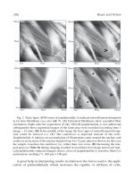
... the recording of pictures with increasing detail. Especially in fluid medium, the investigation of active cells offers numerous facts. As an example of dynamic interactions, a series of images ... magnified in Fig. 6). 116 Bischoff et al. 116 Fig. 6. Zooming in on frame B of Fig. 5 (same z scale for both figures). Increasing contrast in the deflection mode indicates growing roughness of the ... explained in detail in Fig. 11. 7.9. Cells Can Contaminate or Stick to the Scanning Tip Occasionally during the scanning procedure, the tip is covered with an in- definite cluster. Tip-biofouling...
Ngày tải lên: 06/08/2014, 02:20
Atomic Force Microscopy in Cell Biology Episode 1 Part 7 pot
... 135 nominal setpoint used during imaging in the case of curve 3, we have an inden- tation of 950 nm, and for curve 4 we find 200 nm. We need to keep these features and figures in mind in order ... continuously during dem- ineralization in the wet cell of the AFM. Thus, each pixel of a line and each line of a scan represent different exposures and offer a continuous record of the demineraliza- tion ... Bischoff, R., Bischoff, G., and Hein, H J. (2002) Scanning force microscopy (SFM) visualization of adherently growing cells. Am. Biotech. Lab. 3, 20–22. 20. Bischoff, R., Bischoff, G., and Hoffmann,...
Ngày tải lên: 06/08/2014, 02:20
Atomic Force Microscopy in Cell Biology Episode 1 Part 8 pptx
... surrounding intertubular dentin, leaving enlarged tubule lumens (Fig. 4B). Exposure increments are continued in steps of 10 s to a minute or more to follow continued changes in the dentin structure ... ν i 2 )/E i (6) Indentations can be made at intervals of 1–2 µm using indents of submicro- scopic size (approx 300–500 nm). The spacing is needed to avoid influence of one indent on the adjacent indent ... Nanoindentation of Demineralized Dentin There is considerable interest in measuring dentin or other calcified tissues as modified by demineralization or hypomineralization. For example, etching procedures...
Ngày tải lên: 06/08/2014, 02:20
Atomic Force Microscopy in Cell Biology Episode 1 Part 9 docx
... (1995) Atomic force microscopy study of fine structures of the entire surface of red blood cells. Scanning Microscopy 9, 981–988. 5. Siedlecki, C. A. and Marchant, R. E. (1998) Atomic force microscopy ... previous scanning tunneling microscope (5), which provided information at atomic resolution of specimens that are electrically conducing. Because SFMs involve interactions between atomic forces (about ... Adapting atomic force microscopy for cell biology. Ultramicroscopy 82, 289–195. 3. Gunning, P. A., Kirby, A. R., Parker, M. L., Gunning, A. P., and Morris, V. J. (1996) Comparative imaging of Pseudomonas...
Ngày tải lên: 06/08/2014, 02:20
Atomic Force Microscopy in Cell Biology Episode 1 Part 10 potx
... with different information (Fig. 3). As outlined in Fig. 3, applying low imaging forces will result in a more accurate surface visualization (Fig. 3B), whereas increasing the force will result in obscuring ... daptomycin: atomic force microscopy investigation. Chemotherapy 14, 336-341. 14. Nagao, E. and Dvorak, J. A. (1999) Developing the atomic force microscope for studies of living cells. Intern. ... Wisse 1. Introduction In 1986, Binnig et al. (1) revolutionized microscopy through the invention of the atomic force microscope (AFM). Subsequently, commercial instruments of this new imaging technique...
Ngày tải lên: 06/08/2014, 02:20
Atomic Force Microscopy in Cell Biology Episode 2 Part 1 ppsx
... AFM of Protein Complexes 217 217 16 Atomic Force Microscopy of Protein Complexes Olga I. Kiselyova and Igor V. Yaminsky 1. Introduction Scanning probe microscopy (SPM) is a rather new family of ... versa. (D) Increasing the inte- gral gain (see Note 16 during imaging resulted in an artifact-free image and reveal typi- cal jasplakinolide-induced changes, that is, a loss of jasplakinolide A-sensitive ... marked line in (A). AFM Imaging of Living Cells 209 3.5.3. Tip-Induced Smearing In general, the lateral force can wipe away or smear out surface features, whereas the constant force can deform soft...
Ngày tải lên: 06/08/2014, 02:20

Atomic Force Microscopy in Cell Biology Episode 2 Part 2 ppt
... degree of tilt of the molecules in the gel compared with the fluid phase because the depression of the tip in a softer fluid phase varies depending on the composition of this phase. In case of BLES, ... edge of the gel domains (indicated by letter C in the images) in (A) are not smooth but contain differ- ent filamentous structure and microdomain (arrow) protruding from such domains as shown in ... states using a combination of fluorescence and atomic force microscopy (AFM) (9). Monolayer films have also become a standard model for studying lipid– protein interactions and associations in biological...
Ngày tải lên: 06/08/2014, 02:20

Atomic Force Microscopy in Cell Biology Episode 2 Part 3 pps
... (6–9). Since its invention in 1986, the use of the atomic force microscopy (AFM) has become a standard technique on various biological applications, including chromosomes (10–12), not requiring, ... loading force. Do not exceed a loading force of 1 nN. Start with a loading force of 0.5 nN and then switch to the image. When the cantilever is in contact with the cell surface, increase the integral ... part of the force curve and the set-point line. This value (in nm) multiplied by the spring constant gives the loading force (for details, see Appendix). You should not exceed a loading force of...
Ngày tải lên: 06/08/2014, 02:20

Atomic Force Microscopy in Cell Biology Episode 2 Part 4 docx
Ngày tải lên: 06/08/2014, 02:20

Atomic Force Microscopy in Cell Biology Episode 1 Part 1 pdf
Ngày tải lên: 06/08/2014, 02:20

Atomic Force Microscopy in Cell Biology Episode 1 Part 2 pps
Ngày tải lên: 06/08/2014, 02:20

Atomic Force Microscopy in Cell Biology Episode 1 Part 4 docx
Ngày tải lên: 06/08/2014, 02:20

Atomic Force Microscopy in Cell Biology Episode 1 Part 5 docx
Ngày tải lên: 06/08/2014, 02:20