t2 and pd weighted and flair magnetic resonance imaging

Synthesis of superparamagnetic nanostructures and their magnetic resonance imaging applications
... intrinsic and extrinsic effects of field inhomogeneities, it is termed T2* , which is related to T2 by the equation: T2 * T2 ,int T2 ,ext - (Eqn 2) where T2, int is the intrinsic T2 effect, and T2, ext ... nanocluster JCPDS - Joint Committee on Powder Diffraction Standards MFN – Manganese ferrite nanoparticles MFNC – Manganese ferrite nanocomposite MRI – Magnetic resonance imaging MRSI – Magnetic resonance ... Diagnostic Imaging 1 1.2 Magnetic Resonance Imaging 5 1.2.1 T1 Contrast Effect 7 1.2.2 T2 Contrast Effect 8 1.3 Advances in Contrast Agents for Magnetic Resonance...
Ngày tải lên: 09/09/2015, 17:55

Báo cáo y học: "Conventional and diffusion-weighted magnetic resonance imaging findings of benign fibromatous paratesticular tumor: a case report" pptx
... Histopathology reported an Figure T2- weighted images (a) Transverse and (b) sagittal T2- weighted images show tumor heterogeneity The mass (arrow) was mainly hypointense on T2- weighted images, a finding ... of high T2 signal and ADC value of 1.56 × 10⁻³mm2/s, and others of very low T2 signal and ADC value of 0.86 × 10⁻³ mm2/s (Figures 2a, b, 3b) A large, left hydrocele, with a few septa and ADC ... coil The study included fast spin-echo axial, sagittal and coronal T 2weighted sequences and spin-echo axial T1 -weighted sequences Diffusion imaging was performed in the axial plane, using a single...
Ngày tải lên: 11/08/2014, 00:23

Tài liệu Magnetic resonance imaging and gynecological devices doc
... of interaction between magnetic resonance imaging and the copper-T380A IUD Contraception 1997;55:169–73 543 [13] Zieman M, Kanal E Copper T 380A IUD and magnetic resonance imaging Contraception ... or implants Material and methods The authors researched in PubMed-Medline/Ovid using the following keywords: Magnetic Resonance Imaging, Intrauterine Devices, Implanon® and Essure® This article ... superparamagnetic or ferromagnetic) In the presence of discordant (antiparallel) magnetization, the substances are classified as exhibiting negative magnetic susceptibility and are called diamagnetic...
Ngày tải lên: 13/02/2014, 07:20

báo cáo hóa học:" Dynamic magnetic resonance imaging in assessing lung function in adolescent idiopathic scoliosis: a pilot study of comparison before and after posterior spinal fusion" doc
... ultrafast dynamic breath-hold (BH) MR imaging and multiplanar reformat technique, the lung volume, chest wall, and diaphragmatic motions between inspiration and expiration can be accurately measured ... apex and counter pressures at opposite ends under SSEP monitoring Rod estimation and preliminary contouring was then made Decortication of facet joints and transverse processes was made and bone ... into axial and coronal planes so that motions of the chest wall and diaphragm could be assessed The chest wall and diaphragmatic motions were measured in antero-posterior, left-right and cranio-caudal...
Ngày tải lên: 20/06/2014, 01:20

Pocket Atlas of Sectional Anatomy Computed Tomography and Magnetic Resonance Imaging - Volume II Thorax, Heart, Abdomen, and Pelvis (Part 1 ) pptx
... III Pocket Atlas of Sectional Anatomy Computed Tomography and Magnetic Resonance Imaging Volume II Thorax, Heart, Abdomen, and Pelvis Torsten B Moeller, MD Department of Radiology Caritas ... fortunately, constantly evolving Imaging diagnosis is no exception Technical improvements guarantee further developments in diagnosis Computed tomography (CT) and magnetic resonance imaging (MRI) have attained ... processing and storage 12345 Moeller, Sectional Anatomy © 2007 Thieme All rights reserved Usage subject to terms and conditions of license V Dedicated to our parents, Alfred and Friedel Möller and...
Ngày tải lên: 29/06/2014, 09:21
Pocket Atlas of Sectional Anatomy Computed Tomography and Magnetic Resonance Imaging - Volume II Thorax, Heart, Abdomen, and Pelvis (Part 2 ) doc
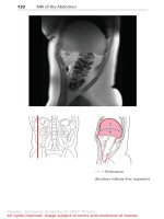
... 31 32 Rib Intercostal artery, vein and nerve Intercostalis muscles Costodiaphragmatic recess Latissimus dorsi muscle Adrenal gland Right kidney Renal artery and vein Ureter Quadratus lumborum ... 22 Diaphragm (lumbar part) 23 Splenic artery and vein 24 Adrenal gland 25 Left kidney 26 Intertransversarii muscles (lateral lumbar) 27 Renal artery and vein 28 Latissimus dorsi muscle 29 Erector ... subject to terms and conditions of license 165 166 MR Angiography of the Splenic and Portal Veins Moeller, Sectional Anatomy © 2007 Thieme All rights reserved Usage subject to terms and conditions...
Ngày tải lên: 29/06/2014, 09:21

Báo cáo y học: "Autologous chondrocyte implantation for cartilage repair: monitoring its success by magnetic resonance imaging and histology" ppt
... O’Driscoll; MRI, magnetic resonance imaging; n/a not applicable; N/A not available with standard and polarised light and images captured and digitised using a closed-circuit television and Image Grabber ... 53 Chung CB, Frank LR, Resnick D: Magnetic resonance imaging: state of the art Cartilage imaging techniques Current clinical applications and state of the art imaging Clin Orthop 2001, 391(suppl):S370-S376 ... proton magnetic resonance microscopy Arthritis Rheum 2000, 43:1580-1590 Recht M, Bobic V, Burstein D, Disler D, Gold G, Gray M, Kramer J, Lang P, McCauley T, Winalski C: Magnetic resonance imaging...
Ngày tải lên: 09/08/2014, 01:21
Báo cáo y học: "Magnetic resonance imaging changes of sacroiliac joints in patients with recent-onset inflammatory back pain: inter-reader reliability and prevalence of abnormalities" potx
... symptoms and the diagnosis of AS is reported [3,4] Such a delay is increasingly unwarranted because of the availability of effective treatment Magnetic resonance imaging (MRI) is an imaging modality ... reliability and sensitivity to change over one year J Rheumatol 1999, 26:997-1002 Braun J, Bollow M, Eggens U, Konig H, Distler A, Sieper J: Use of dynamic magnetic resonance imaging with fast imaging ... changes and structural changes and the different localization of these changes Pathological changes of interest were defined as inflammation and structural changes including erosions, sclerosis and...
Ngày tải lên: 09/08/2014, 07:20

Báo cáo y học: "Long term evaluation of disease progression through the quantitative magnetic resonance imaging of symptomatic knee osteoarthritis patients: correlation with clinical symptoms and radiographic change" pps
... of at least 24 months and a cohort of several thousands is necessary to establish the effect of pharmacological interventions on OA disease progression Magnetic resonance imaging (MRI) allows ... JPR, JMP, JMM, JFB, GAC, and JPP analyzed and interpreted the data JPR, JMP and JPP prepared the manuscript JPR and GAC performed the statistical analysis All authors read and approved the final ... M, et al.: Reliability of a quantification imaging system using magnetic resonance images to measure cartilage thickness and volume in human normal and osteoarthritic knees Osteoarthritis Cartilage...
Ngày tải lên: 09/08/2014, 07:20

Báo cáo y học: "proximal interphalangeal joints in rheumatoid arthritis: a comparison with magnetic resonance imaging, conventional radiography and clinical examination" pdf
... synovitis (grade 4) Axial T1 -weighted magnetic resonance images were obtained (b) before and (c) after contrast administration (grade synovitis) MRI, magnetic resonance imaging; RA, rheumatoid arthritis ... longitudinal and (b) the transverse planes shows both signs of destruction (grade 2) and inflammation (grade 3) Axial T 1weighted magnetic resonance images were obtained (c) before and (d) after ... Additionally, a coronal T1 -weighted magnetic resonance image (e) before contrast administration visualizes the same bone erosion as shown in panels c and d The coronal magnetic resonance image of the...
Ngày tải lên: 09/08/2014, 07:20

Báo cáo y học: "Are bone erosions detected by magnetic resonance imaging and ultrasonography true erosions? A comparison with computed tomography in rheumatoid arthritis metacarpophalangeal joints" pptx
... ultrasound, and contrast-enhanced magnetic resonance imaging Arthritis Rheum 1999, 42:1232-1245 Klarlund M, Østergaard M, Jensen KE, Madsen JL, Skjødt H, Lorenzen I: Magnetic resonance imaging, ... radiographs Imaging evaluation All imaging modalities were evaluated with investigators blinded to clinical and other imaging data Each MCP joint quadrant (radial and ulnar part of the metacarpal head and ... CH, Lorenzen I: Magnetic resonance imaging- determined synovial membrane and joint effusion volumes in rheumatoid arthritis and osteoarthritis: comparison with the macroscopic and microscopic...
Ngày tải lên: 09/08/2014, 08:22
Báo cáo y học: "Correlation of histopathological findings and magnetic resonance imaging in the spine of patients with ankylosing spondylitis" potx
... correlation of bone marrow edema in histopathological assessment and magnetic resonance imaging (a) Magnetic resonance imaging (T2 imaging sequence) of a zygapophyseal joint with bone marrow edema ... correlation of bone marrow edema in histopathological assessment and magnetic resonance imaging (a) Magnetic resonance imaging of imaging zygapophyseal joints (cyan arrows) without detectable bone ... four micro-vessels per HPF; ++, five or six micro-vessels per HPF Magnetic resonance imaging MRI, including T1 -weighted, T2- weighted and TIRM (turbo inversion recovery magnitude) sequences, was performed...
Ngày tải lên: 09/08/2014, 08:22

Báo cáo khoa học: " Comparison of T2 and FLAIR imaging for target delineation in high grade gliomas" pptx
... created from the union of the T2 and FLAIR CTVs with a standard cm volumetric expansion The percent difference and absolute difference between the combined PTV and the T2 and FLAIR PTV was calculated ... dose of 60 Gy T2 and FLAIR volumes There was a large range in the size of CTVs and PTVs across the patient cohort The mean T2 and FLAIR CTVs were 98.99 cc (range 1.51-383.5 cc) and 113.76 cc ... combined PTV and the T2 and FLAIR PTVs was 46.23 cc and 18.67 cc Using the combined PTV would result in an 11.7% variation from the PTV as defined by T2 and 4.16% as defined by FLAIR 20 Normal...
Ngày tải lên: 09/08/2014, 10:20

Báo cáo y học: "The association between patellar alignment on magnetic resonance imaging and radiographic manifestations of knee osteoarthritis" ppsx
... a field of view (FOV) of 11 to 12 cm, and a matrix of 256 pixels × 192 pixels; and coronal and axial spinecho fat-suppressed proton density -weighted and T 2weighted images (TR 2,200 ms; TE 20/80 ... device was used to ensure uniformity between patients The imaging protocol included sagittal spin-echo proton density -weighted and T2- weighted images (repetition time (TR) 2,200 ms; time to echo ... by magnetic resonance imaging (MRI) [19-21] Muellner and colleagues [19] performed measurements analogous to those used in Xray evaluation with MRI images obtained with knees flexed to 20° and...
Ngày tải lên: 09/08/2014, 10:20

Báo cáo y học: "Cartilage markers and their association with cartilage loss on magnetic resonance imaging in knee osteoarthritis: the Boston Osteoarthritis Knee Study" doc
... view (FOV) of 11 to 12 cm, and a matrix of 256 × 192 pixels and coronal and axial spin-echo fat-suppressed proton density- and T 2weighted images (TR 2,200 milliseconds and TE 20/80 milliseconds) ... Center and the Veterans Administration Boston Health Care System approved the baseline and follow-up examinations, and informed consent was obtained from all participants Magnetic resonance imaging ... excitation, and the same FOV and matrix Cartilage on MRI was scored paired and unblinded to sequence on 14 plates (anterior, central, and posterior femur; anterior, central, and posterior tibia; and...
Ngày tải lên: 09/08/2014, 10:21
Báo cáo y học: "Ultrasonography, magnetic resonance imaging, radiography, and clinical assessment of inflammatory and destructive changes in fingers and toes of patients with psoriatic arthritis" pot
... and/ or effusion, and/ or power Doppler cmagnetic resonance imaging (MRI) synovitis Values are absolute agreement presented as a bclinically signal); tender and swollen joints; percentage The imaging ... intratendinous PD signal at the entheses Ultrasonography parameters The setting for grey-scale US was 14 MHz, and the pulse repetition frequency for the PD signal was set at 500 Hz Magnetic resonance imaging ... radiography (MH), who was blinded to clinical and other imaging findings Magnetic resonance imaging parameters The acquired images included a coronal T1 -weighted threedimensional fast field echo...
Ngày tải lên: 09/08/2014, 10:21
Báo cáo y học: "Detection of bone erosions in rheumatoid arthritis wrist joints with magnetic resonance imaging, computed tomography and radiography" ppt
... tomography and (c, d) T1patient weighted magnetic resonance imaging in the (a, c) coronal and (b, d) axial planes A bone erosion at the distal radius is seen on both computed tomography and magnetic resonance ... Computerized measurement of magnetic resonance imaging erosion volumes in patients with rheumatoid arthritis: a comparison with existing magnetic resonance imaging scoring systems and standard clinical outcome ... ultrasound, and contrast-enhanced magnetic resonance imaging Arthritis Rheum 1999, 42:1232-1245 Klarlund M, Østergaard M, Jensen KE, Madsen JL, Skjødt H, Lorenzen I: Magnetic resonance imaging, ...
Ngày tải lên: 09/08/2014, 10:23

báo cáo khoa học: " N-hexanoyl chitosan stabilized magnetic nanoparticles: Implication for cellular labeling and magnetic resonance imaging" pps
... longitudinal T2 weighted T2 weighted MR images of a representive RAW cells incubated with different volume of MC-IOPs (11.2 mg/ml) for h, (a) longitudinal section, (b) coronal section and (c) signal ... (curve c), and MCIOPs (curve d) IOPs exhibit strong bands in the low frequency region below 800 cm-1 due to the oxide skeleton The characteristic bands of modified chitosan, amide I, II, and III ... spectra of IOPs have weak bands The spectrum is consistent with magnetic (Fe3O4) and the signals associated to the magnetite appear as broad features at 408.9, 571.5 and 584.5 cm-1 [4] Figure...
Ngày tải lên: 11/08/2014, 00:22

báo cáo khoa học: "Optimized labeling of bone marrow mesenchymal cells with superparamagnetic iron oxide nanoparticles and in vivo visualization by magnetic resonance imaging" ppsx
... high-resolution magnetic resonance imaging Biophys J 1999, 76:103-109 Jendelova P, Herynek V, Urdzikova L, Glogarova K, Kroupova J, Andersson B, Bryja V, Burian M, Hajek M, Sykova E: Magnetic resonance ... superparamagnetic iron oxide nanoparticles and in vivo visualization by magnetic resonance imaging Journal of Nanobiotechnology 2011 9:4 Submit your next manuscript to BioMed Central and take ... [11-13] Magnetic resonance imaging (MRI) is an excellent tool for high-resolution visualization of the fate of cells after transplantation and for evaluation of cell-based repair, replacement, and...
Ngày tải lên: 11/08/2014, 00:22
