fiber x ray diffraction and dna double helix

High resolution x ray diffraction study of phase and domain structures and thermally induced phase transformations in PZN (4 5 9)%PT 1
... carbide XRD : x- ray diffraction HR-XRD : High-resolution synchrotron x- ray diffraction RSM : Reciprocal space mapping PLM : Polarized light microscope xx FWHM : Full-width-at-half-maximum ε’ ... et al.) S: x = ([001]-poled) S: x = ([001]- and [111]-poled) S: x = 5, (unpoled) S: x =4.5, ([001]-poled) S: x = 4.5, ([001]-poled) S: x =8, 10 ([100]-,[011]-, and [111]-poled) P: 10 x 15 prepared ... PZN-PT PZN -x% PT: single crystal (S) or powder (P) P: 5
Ngày tải lên: 14/09/2015, 08:40
High resolution x ray diffraction study of phase and domain structures and thermally induced phase transformations in PZN (4 5 9)%PT 2
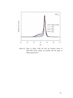
... Synchrotron x- ray diffraction of higher energy, being 18 keV, revealed similar penetration depth in PZN-8%PT sample [32] Meanwhile, using synchrotron x- ray diffraction at 10.7 keV, 32 keV, and 67 ... µm, 59 µm, and 412 µm of a PZN sample [57] This suggests a nonlinear relation between the x- ray energy and the penetration depth despite similar test samples were used Whether the x- ray diffraction ... surface X- ray 44.65o ~43.0-44.0o 42 43 44 45 46 47 2θ [010]W [100] L PZN-4.5%PT b) b) Fractured surface Intensity (arb units) X- ray X- ray 44.62 42 43 44 45 o 46 47 2θ Figure 5.1 (002) XRD profiles...
Ngày tải lên: 14/09/2015, 08:40

High resolution x ray diffraction study of phase and domain structures and thermally induced phase transformations in PZN (4 5 9)%PT 3
... PZN-8%PT, and PZN-9%PT single crystals The results are provided in Figures 5.11(a), (c), and (e) To expose the bulk material, the same samples were later fractured into two halves and were again x- rayed ... 5.11(b), (d), and (f) The polished -and- annealed bulk PZN-PT single crystals were found to cover with a deformed layer even after heating to above TC, giving rise to smear effect in the x- ray diffraction ... occurrence of the extremely broad lower 2θ peak adjacent to the main (002)R peak in standard XRD profiles of (001)-orientated PZN-PT single crystals has been examined This diffraction arises...
Ngày tải lên: 14/09/2015, 08:40
High resolution x ray diffraction study of phase and domain structures and thermally induced phase transformations in PZN (4 5 9)%PT 4
... in the x- ray profile following any direction of polishing, suggesting that the surface phase is always present regardless of the direction and/ or mode of polishing This behavior can be explained ... annealing treatments The heating and cooling rates used were 1.5 ºC/min Figure 5.9 shows the (002) XRD profiles of the differently annealed PZN-4.5%PT samples and the (002) XRD profile of the as-polished ... 5.7a), was tilted by an angle of 45° and again polished back and forth in the new direction After the second polishing, the earlier set of domains disappeared and was replaced by a new set of elongated...
Ngày tải lên: 14/09/2015, 08:40

High resolution x ray diffraction study of phase and domain structures and thermally induced phase transformations in PZN (4 5 9)%PT 5
... the diffraction 88 patterns may be assigned as: (a) MC (d1, d3, d7, d4, and d6, d7 being the bm diffraction) + T (d2 and d5), (b) T (d2 and d5) + T* (d1, d3, d4 and d6) + R (d7), or (c) T (d2 and ... micro- and nanodomains and Figures 6.2(d)-(h) are the corresponding diffraction patterns on the (002) RSM To explain how domain size may affect the (002) diffraction profiles, we begin with diffractions ... parameters am and cm are equal Because reciprocal lattice points in HR-XRD RSM are projected with respect to the pc axes, it is important to establish the relationship between the pc axes and the axes...
Ngày tải lên: 14/09/2015, 08:40

High resolution x ray diffraction study of phase and domain structures and thermally induced phase transformations in PZN (4 5 9)%PT 6
... of coexistence of the {100}-type and {110}-type R nanotwins onto the (002) RSM (b) The projection of the coexistence of R micro- and nanotwins onto (002) RSM 112 coexistence of R micro- and nanotwin ... 7.2(a) To understand how the (002) diffraction pattern is affected by: (a) the four structural variants of the R structure, i.e., r1, r2, r3, and r4, and (b) its domain size, the diffraction produced ... {100}-type and/ or {110}-type R microtwin domains, a result of the large diffraction width associated with the fine domain structure and the extreme compliant nature of the R phase The diffraction...
Ngày tải lên: 14/09/2015, 08:40

High resolution x ray diffraction study of phase and domain structures and thermally induced phase transformations in PZN (4 5 9)%PT 7
... the cooling and transformation stresses to be relaxed via twinning of both the untransformed T and the transformed R phases Thus, at room condition, the mixture of R and T micro- and/ or nanotwins ... unpoled (annealed) (a) and (b) PZN-7%PT, and (c) and (d) PZN-8%PT single crystals 127 sample of PZN-7%PT and PZN-8%PT, respectively, but of the same wafers as in Figures 8.1(a) and (c) These peaks ... 6.1 gives the relationships between the m axes and the pc axes of the various M phases and the O phase Judging from the nature of splitting, these diffraction peaks in Figure 8.1(d), say, may...
Ngày tải lên: 14/09/2015, 08:40

High resolution x ray diffraction study of phase and domain structures and thermally induced phase transformations in PZN (4 5 9)%PT 8
... 8.13e) may be assigned as (a) MB (d1, d3, d4, and d6) + T (d2 and d5), (b) MC (d1, d2, d3, d4, and d6) + T (d2 and d5) or T* (d1, d3, d4, and d6) + T (d2 and d5) (see Section 6.2.3 for details) The ... to Tmax ≅ 165 ºC, the scattered areas started to exhibit extinction at most P/A angles, suggesting the occurrence of the C phase (Figure 8.14f) Above Tmax, the sample exhibited a total extinction ... (d1, d2, d3, d4, and d6) + T (d2 and d5), or (c) T* (i.e., d1, d3, d4, and d6) + T (i.e., d2 and d5) We may rule out possibilities (a) and (b) as transformations of the type MB-C and MC-C are not...
Ngày tải lên: 14/09/2015, 08:40

High resolution x ray diffraction study of phase and domain structures and thermally induced phase transformations in PZN (4 5 9)%PT 9
... the entire MPB (standard + extended) region where (R+T) coexist, the crystal exhibits extremely high piezoelectric properties This suggests that the presence of (R+T) phase mixture in the form ... nanotwins if the diffraction is of considerably smaller FWHM and lies in the ω = 0°, or (c) the convoluted peak of both {100}-type and {110}-type R micro- and nanotwin mixture if the diffraction is ... structure Samples showing similar RSMs were ground into powder of
Ngày tải lên: 14/09/2015, 08:40

High resolution x ray diffraction study of phase and domain structures and thermally induced phase transformations in PZN (4 5 9)%PT 10
... current and HR-XRD results suggest TR-T ≅ 100-135 °C for [001]-annealed -and- poled PZN-4.5%PT and 95-115 °C for [001]-annealed-andpoled PZN-7%PT single crystals 175 Table 9.2 TDP, TR-T(L), and TR-T(U) ... transformation in relaxor single crystals occurs over a temperature range, manifested by the coexistence of R* and T* domains, a string of thermal current signals, and continued degradation of KT and k31 The ... the x- axis Both the KT and k31 of PZN-4.5%PT started to degrade after it was heated to 105 ºC 174 domains are parallel to the E-field direction which is normal to the fractured surface being x- rayed...
Ngày tải lên: 14/09/2015, 08:40

High resolution x ray diffraction study of phase and domain structures and thermally induced phase transformations in PZN (4 5 9)%PT 11
... the ferroelectric relaxor (1 -x) Pb(Mg1/3Nb2/3)O3-xPbTiO3, Phys Rev B, 66, p 054104 2002 36 Bai, F., N Wang, J Li, D Viehland, P M Gehring, G Xu and G Shirane X- ray and neutron diffraction investigations ... Bellaiche, L., A Garcia and D Vanderbilt Electric-field induced polarization paths in Pb(Zr1-xTix)O3 alloys, Phys Rev B, 64, p 060103 2001 25 Vanderbilt, D and M H Cohen Monoclinic and triclinic phases ... to an expanded MPB across 0.06 ≤ x ≤ 0.10 in PZN-xPT single crystals This expanded (R+T) MPB region can be further divided into two different regions In the low PT region (i.e., 0.06 ≤ x ≤ ≈ 0.08),...
Ngày tải lên: 14/09/2015, 08:40

Tài liệu Báo cáo khoa học: X-ray crystallographic and NMR studies of pantothenate synthetase provide insights into the mechanism of homotropic inhibition by pantoate docx
... Pro59–Gln61 and Glu119–Ser122 that are present in the crystal structures of E coli and M tuberculosis PS, are missing in the solution structure Helix IX, which is a continuous helix (16 residues) ... backbone and side chain 13Ca and 13C¢ and 13Cb carbons are assigned to the extent of 97%, 93% and 93%, respectively (BMRB accession number 6940) [24] The H–15N correlation in the TROSY experiment ... form of nPS using X- ray crystallography, as described below A Solution and refinement of the structure using X- ray crystallography B The crystal structure of the pantoate–nPS complex was solved by...
Ngày tải lên: 16/02/2014, 09:20

Tài liệu Báo cáo khoa học: Molecular determinants of ligand specificity in family 11 carbohydrate binding modules – an NMR, X-ray crystallography and computational chemistry approach doc
... when the complex is formed The ligands cellobiose, cellotetraose and cellohexaose were studied Results and Discussion The crystal structure of CtCBM11, the binding cleft and its ligand specificity ... in the X- ray structures of CBM4 and CBM17 complexed with cellopentaose and cellohexaose, respectively [13,23] The involvement of the tyrosine residues in the stabilization of the complex cannot ... two six-stranded anti-parallel b-sheets that form a convex side (b-strands 1, 3, 4, 6, and 12) and a concave side (b-strands 2, 5, 7, 8, 10 and 11) The concave side is decorated by the side chains...
Ngày tải lên: 18/02/2014, 17:20
Báo cáo khoa học: The effect of replacing the axial methionine ligand with a lysine residue in cytochrome c-550 from Paracoccus versutus assessed by X-ray crystallography and unfolding ppt
... weeks and repeated dissolving of the crystalline networks a single dark red crystal suitable for X- ray diffraction was obtained grown from 0.1 m bicine pH 9.0 and 3.2 m ammonium sulfate X- ray diffraction ... substituents are extremely sensitive to the chemical nature of the axial ligands to the hemeiron [41] Upon replacing the native Met ligand with either an exogenous or protein-based ligand a change ... structure and unfolding upon replacing the axial Met ligand with a Lys in the M100K variant of cyt c-550 from P versutus We describe three X- ray structures, one of the ferric wild type (wt) and two...
Ngày tải lên: 07/03/2014, 17:20
Báo cáo Y học: Determinants of the inhibition of a Taiwan habu venom metalloproteinase by its endogenous inhibitors revealed by X-ray crystallography and synthetic inhibitor analogues pdf
... superfamily of metzincin which exhibits some typical structural features, such as the Met-turn and active-site consensus HExxHxxGxxH sequence [15–17] Some organisms and mammalian tissues recently ... Huber, R & Bode, W (1995) X- ray structures of human neutrophil collagenase complexed with peptide hydroxamate and peptide thiol inhibitors Implications for substrate binding and rational drug design ... off by a solvent mixture of trifluoroacetic acid and ethanedithiol, and solvent was evaporated to dryness The resins were then washed with cold ether and the peptides were extracted with 5% acetic...
Ngày tải lên: 08/03/2014, 23:20
Báo cáo khoa học: An X-ray diffraction study of model membrane raft structures doc
... Supporting information Synchrotron X- ray diffraction measurements X- Ray diffraction measurements were performed on beamline 2.1 at the Daresbury Laboratory The X- ray wavelength was 0.154 nm with ... Northampton, MA, USA) Analysis of X- ray diffraction data The small-angle X- ray scattering-intensity profiles were analysed using standard procedures [47] Polarization and geometric corrections for ... of each phospholipid was performed by ESI-tandem MS [29] and the data are presented in Table S1 4694 Sample preparation Samples for X- ray diffraction examination (Table S2) were prepared by dissolving...
Ngày tải lên: 15/03/2014, 23:20
Báo cáo Y học: Boar salivary lipocalin Three-dimensional X-ray structure and androstenol/androstenone docking simulations pot
... Docking experiment with androstenol and androstenone The previously identified natural ligands androstenol and androstenone have been built with the program ACD/CHEMSKETCH [18] The parameter and topology ... Stereoview of SAL X- ray structure in ribbon representation The helix is in red, strands are blue and turns or coil are yellow The GlcNAc residue bound to Asn53 is in white ball -and- stick, and the serendipitous ... the model of the complex of androstenol bound in SAL, with in (A) and (C) the ligand in position ÔinÕ, with the OH pointing inside the cavity and, in (B) and (D) the ligand in position ÔoutÕ,...
Ngày tải lên: 17/03/2014, 17:20
Báo cáo khoa học: X-ray crystallography and structural stability of digestive lysozyme from cow stomach doc
... residues Glu35 and Asp52 (numbering for HEWL), for BSL2 and other lysozymes, with propka 2.0 [16] The predicted pKa values were 6.15 and 4.27 for BSL2, 5.93 and 4.20 for HEWL, and 4.89 and 3.84 for ... acetate (for pH and 5) and sodium phosphate (for pH and 7) buffer The ionic strength of each buffer was adjusted to 0.1 [40] Lysozyme solution and M lysodeikticus suspension were mixed, and the decrease ... recombinant bovine stomach lysozyme (BSL2), the most highly expressed lysozyme in the cow stomach X- ray crystallography and some other experiments were performed to determine how this lysozyme has...
Ngày tải lên: 23/03/2014, 04:21
Báo cáo khoa học: Structural flexibility in Trypanosoma brucei enolase revealed by X-ray crystallography and molecular dynamics pdf
... 20 mm of ligand (PEP, PAH or FPAH) for and flash-cooled X- ray diffraction data were collected from two native crystals (referred to as sulfate_2 and sulfate_3 in Table 1) and four ligand-cocrystallised ... LiOH provided the expected products PAH and FPAH as lithium salts in 28% and 15% overall yield, respectively X- ray crystallography Recombinant T brucei enolase was expressed and purified as previously ... sulfate_2, PEP and PAH_1 structures is shown in Fig Unexpected variability in inhibitor complex structures Comparison of the substrate and inhibitor complexes shows that the His156-out and His156-in...
Ngày tải lên: 23/03/2014, 07:20

ELEMENTS OF X-RAY DIFFRACTION ppt
... angstrom, the exact relation The region between netic X X X bemg lkX= 1.00202A worth while to review briefly some properties of electromagnetic Suppose a monochromatic beam of x- rays, i.e., x- rays of ... E, (a) with t at a fixed value of x and (b) with x at PKOPERTIES OF X- RAYS [CHAP film arbitrary units, such as the degree of blackening of a photographic to the x- ray beam exposed An accelerated ... high -power x- rav tubes FIG 1-17 Since an x- ray tube is less than percent efficient in producing x- rays since the diffraction of x- rays by crystals is far less efficient than this, and it follows...
Ngày tải lên: 29/03/2014, 11:20