disorders of the eye ppt

Chapter 029. Disorders of the Eye (Part 12) ppt
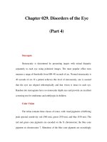
... common in people of northern European descent. Their diagnosis is obvious when they are visible as glittering particles upon the surface of the optic disc. However, in many patients they are hidden ... fulminant papilledema. Optic Disc Drusen These are refractile deposits within the substance of the optic nerve head (Fig. 29-13). They are unrelated to drusen of the retina, which occur in age-related ... beneath the surface, producing pseudo-papilledema. It is important to recognize optic disc drusen to avoid an unnecessary evaluation for papilledema. Chapter 029. Disorders of the Eye (Part...
Ngày tải lên: 06/07/2014, 15:21

Chapter 029. Disorders of the Eye (Part 18) ppt
... limitation of motility. The width of the palpebral fissures is measured in primary gaze to quantitate the degree of ptosis. The ptosis will be underestimated if the patient compensates by lifting the ... weakness), or a family history of ptosis should be sought. Fluctuating ptosis that worsens late in the day is typical of myasthenia gravis. Chapter 029. Disorders of the Eye (Part 18) Orbital ... drooping of the eyelid. Unilateral or bilateral ptosis can be congenital, from dysgenesis of the levator palpebrae superioris, or from abnormal insertion of its aponeurosis into the eyelid....
Ngày tải lên: 06/07/2014, 15:21

Chapter 029. Disorders of the Eye (Part 20) ppt
... when the oculomotor nerve is injured by trauma or compression (tumor, aneurysm). Miswiring of sprouting fibers to the levator muscle and the rectus muscles results in elevation of the eyelid ... thought to result from microvascular infarction of the nerve, somewhere along its course from the brainstem to the orbit. Usually the patient complains of pain. Diabetes, hypertension, and vascular ... the subarachnoid space the oculomotor nerve is vulnerable to aneurysm, meningitis, tumor, infarction, and compression. In cerebral herniation the nerve becomes trapped between the edge of the...
Ngày tải lên: 06/07/2014, 15:21

Chapter 029. Disorders of the Eye (Part 23) pptx
... examination of the eyes. Observation of nystagmoid movements of the optic disc on ophthalmoscopy is a sensitive way to detect subtle nystagmus. Gaze-Evoked Nystagmus This is the most common form of ... JJ: Systemic Diseases and the Eye. St Louis, Mosby, 2001 Leibowitz HM: The red eye. N Engl J Med 343:345, 2000 [PMID: 10922425] Leigh RJ, Zee DS: The Neurology of Eye Movements , 4th ed. Oxford, ... lesions, the nystagmus does not appear until several months of age. Congenital motor nystagmus, which looks similar to congenital sensory nystagmus, develops in the absence of any abnormality of the...
Ngày tải lên: 06/07/2014, 15:21

Chapter 029. Disorders of the Eye (Part 14) pps
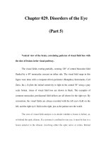
... Chapter 029. Disorders of the Eye (Part 14) Stroke This occurs when interruption of blood supply from the posterior cerebral artery to the visual cortex is prolonged. The only finding ... viewing light reflected from the fundus with an ophthalmoscope or by examining the dilated eye using the slit lamp. The only treatment for cataract is surgical extraction of the opacified lens. Over ... cataracts develop slowly as a result of aging, leading to gradual impairment of vision. The formation of cataract occurs more rapidly in patients with a history of ocular trauma, uveitis, or diabetes...
Ngày tải lên: 06/07/2014, 15:21

Chapter 029. Disorders of the Eye (Part 16) pps
... by administration of panretinal laser photocoagulation at the appropriate point in the evolution of the disease. For further discussion of the manifestations and management of diabetic retinopathy, ... detachment of the retinal pigment epithelium and the neurosensory retina. These detachments produce acute or chronic symptoms of metamorphopsia and blurred vision when the macula is involved. They ... Chapter 029. Disorders of the Eye (Part 16) Central Serous Chorioretinopathy This primarily affects males between the ages of 20 and 50. Leakage of serous fluid from the choroid causes...
Ngày tải lên: 06/07/2014, 15:21

Chapter 029. Disorders of the Eye (Part 17) potx
... 029. Disorders of the Eye (Part 17) Melanoma and Other Tumors Melanoma is the most common primary tumor of the eye (Fig. 29-18). It causes photopsia, an enlarging scotoma, and loss of vision. ... atrophy of retrobulbar fat, or fracture of the orbital floor. The position of the eyes within the orbits is measured using a Hertel exophthalmometer, a hand-held instrument that records the position ... to a therapeutic trial of systemic glucocorticoids indirectly provides the best confirmation of the diagnosis. When the globes appear asymmetric, the clinician must first decide which eye...
Ngày tải lên: 06/07/2014, 15:21

Chapter 029. Disorders of the Eye (Part 19) ppsx
... "lazy" eye) in the deviated eye. Chapter 029. Disorders of the Eye (Part 19) Myogenic Ptosis The causes of myogenic ptosis include myasthenia gravis (Chap. 381) and a number of rare myopathies ... gaze, and then with the head turned and tilted in each direction. In the above example, a cover test with the head turned to the right will maximize the fixation shift evoked by the cover test. ... If the eye movements are full and the ocular misalignment is equal in all directions of gaze (concomitant deviation), the diagnosis is strabismus. In this condition, which affects about 1% of...
Ngày tải lên: 06/07/2014, 15:21

Chapter 029. Disorders of the Eye (Part 21) pps
... generally have the opposite effect: the eyes deviate conjugately away from the irritative focus. Parietal lesions disrupt smooth pursuit of targets moving toward the side of the lesion. Bilateral ... injury is similar, except for the eye findings. There is lateral rectus weakness only, instead of gaze palsy, because the abducens fascicle is injured rather than the nucleus. Infarct, tumor, ... sclerosis are the most common etiologies of brainstem abducens palsy. After leaving the ventral pons, the abducens nerve runs forward along the clivus to pierce the dura at the petrous apex,...
Ngày tải lên: 06/07/2014, 15:21

Chapter 029. Disorders of the Eye (Part 22) ppsx
... combined lesion of the medial longitudinal fasciculus and the abducens nucleus on the same side. The patient's only horizontal eye movement is abduction of the eye on the other side. Figure ... position of gaze the eyes appear normal. B. Horizontal gaze to the left is intact. C. On attempted horizontal gaze to the right, the left eye fails to adduct. In mildly affected patients the eye ... controlled at the level of the midbrain. The neuronal circuits affected in disorders of vertical gaze are not fully elucidated, but lesions of the rostral interstitial nucleus of the medial longitudinal...
Ngày tải lên: 06/07/2014, 15:21
Chapter 029. Disorders of the Eye (Part 8) ppsx
... Chapter 029. Disorders of the Eye (Part 8) Episcleritis This is an inflammation of the episclera, a thin layer of connective tissue between the conjunctiva and sclera. Episcleritis ... cause of a red, painful eye. Susceptible eyes have a shallow anterior chamber, either because the eye has a short axial length (hyperopia) or a lens enlarged by the gradual development of cataract. ... judicious use of topical glucocorticoids. Dilation of the pupil reduces pain and prevents the formation of synechiae. Posterior Uveitis This is diagnosed by observing inflammation of the vitreous,...
Ngày tải lên: 06/07/2014, 15:21


Bạn có muốn tìm thêm với từ khóa: