ch 5 diseases and disorders of the eye and ear

WHEN THINGS GO WRONG – DISEASES AND DISORDERS OF THE HUMAN BRAIN docx
... of our grief, pain, anxiety and tears, is none other than the brain It is specially the organ which enables us to think, see and hear, and to distinguish the ugly and the beautiful, the bad and ... and the good, pleasant and unpleasant It is the brain too which is the seat of madness and delirium, of the fears and frights which assail us” (Taken from Chadwick and Mann’s translation, The ... WRONG – DISEASES AND DISORDERS OF THE HUMAN BRAIN Edited by Theo Mantamadiotis When Things Go Wrong – Diseases and Disorders of the Human Brain Edited by Theo Mantamadiotis Published by InTech Janeza
Ngày tải lên: 28/06/2014, 11:20

Tài liệu Báo cáo khoa học: Treatment of neutral glycosphingolipid lysosomal storage diseases via inhibition of the ABC drug transporter, MDR1 Cyclosporin A can lower serum and liver globotriaosyl ceramide levels in the Fabry mouse model doc
... disease, the primary pathology is in the kidney [1], the major site of Gb3 synthesis in man [11], and in the heart, possibly due to the association of Gb3 synthesis with the microvasculature GSL synthesis ... v) per 100 lL of plasma and then partitioned against ⁄ volume of water The lower phase was dried under a stream of nitrogen gas and then the residue was dissolved in chloroform The sample was ... the Fabry mouse model Our studies have further delineated the abnormal Gb3 synthesis in this model and have shown that the inhibition of MDR1 is a viable potential approach to the reduction of
Ngày tải lên: 19/02/2014, 07:20

báo cáo hóa học:" The reliability, validity, and preliminary responsiveness of the Eye Allergy Patient Impact Questionnaire (EAPIQ)" docx
... deleting either the 'intensity of bother' or 'frequency of bother' items, they were combined in the scoring. The 'intensity of bother' and 'frequency of bother' ... 0.0 056 5 0.69426 0.2 256 9 0.12197 3 Red eyes -0.09104 0.14292 0.80942 0.08124 2 Water eyes 0.10791 0.27184 0 .56 104 -0.13643 1 Swollen / puffy 0.01604 0.36873 0 .54 369 -0.1 055 4 5 Dry eyes 0.492 05 -0.24760 ... presented as posters [4 ,5] . The objective of the present study was to further validate the questionnaire by investigating the psy- chometric properties of the EAPIQ in a sample of patients with ocular
Ngày tải lên: 20/06/2014, 15:20

DISORDERS OF THE VULVA AND VAGINA pps
... therapeutic... and arises in the labia (major and minor) in 65% of patients CHAPTER 20 DISORDERS OF THE VULVA AND VAGINA 59 1 and in the clitoris in 25% Over one third of tumors ... occurs. There may be both erythema and edema of the vulva and vagina. The discharge with Candida infection has a pH of 4? ?5. Mixing the secretions with a drop of 10%–20% KOH microscopically reveals the ... of metastases and subsequent...CHAPTER 20 DISORDERS OF THE VULVA AND VAGINA 58 1 cellulitis, and suppuration of the groin Involvement of apocrine glands establishes the
Ngày tải lên: 05/08/2014, 16:20

ANATOMY, PHYSIOLOGY, AND DISORDERS OF THE AUDITORY SYSTEM - PART 1 pps
... the function of the cochlea than of any other sensory organ C H A P T E R 1 Anatomy of the Ear 1 ABSTRACT 10 1 The ear consists of the outer ear, the middle ear and ... of three parts: the outer ear; the middle ear; and the inner ear The inner ear consists of two parts: the vestibular apparatus for balance; and the cochlea for hearing The ... and the cochlear nerve The smaller sketches show differences between the mammalian cochlea and the avian organs of hearing (reprinted... that disorders of one part of the
Ngày tải lên: 11/08/2014, 06:21

ANATOMY, PHYSIOLOGY, AND DISORDERS OF THE AUDITORY SYSTEM - PART 2 ppsx
... measured in the place of the head. The effect of the head on the sound at the entrance of the ear canal is related to the size of the head and, the wavelength 2 of sound. This means that the “amplification” ... ear by an earphone may reach the other ear by bone conduction (cross transmission). CHAPTER 2 Sound Conduction to the Cochlea 3. HEAD, OUTER EAR AND EAR CANAL The ear canal, the pinna and the ... effect of the head on the sound pressure at the entrance of the ear canal depends on the frequency of the sound and on the angle of incidence of the sound (direction to the sound source). 6. The
Ngày tải lên: 11/08/2014, 06:21
ANATOMY, PHYSIOLOGY, AND DISORDERS OF THE AUDITORY SYSTEM - PART 4 potx
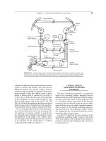
... to the surface of the floor of the fourth ventricle (Fig. 5. 15B) [72]. The other part of the olivocochlear system projects mainly to the contralateral cochlea and the fibers of that system travel ... [76]. The most central part of the corticofugal system originates in the auditory cerebral cortex (Fig. 5. 14A) and the cortico-cochlear system projects from the auditory cortex to the cochlear Chapter ... understanding of the connections in these pathways. Chapter 5 Anatomy of the Auditory Nervous System 85 FIGURE 5. 8 Schematic diagram of the ascending auditory pathways from the left cochlea showing
Ngày tải lên: 11/08/2014, 06:21
ANATOMY, PHYSIOLOGY, AND DISORDERS OF THE AUDITORY SYSTEM - PART 6 pot
... such as the cat, is approximately 0.8 cm long [59 ]. The difference between the latency of the N 1 of the AP and that of the response from the intracranial portion of the auditory nerve is the ... floor of the lateral recess is the (dorsal) surface of the dorsal cochlear nucleus and the rostral portion of the floor of the lateral recess is the dorsal surface of the ... of many skeletal muscles The movement of the eyes toward the source of. .. have shown that the auditory nerve and the ventral cochlear nucleus are the first part of the
Ngày tải lên: 11/08/2014, 06:21
ANATOMY, PHYSIOLOGY, AND DISORDERS OF THE AUDITORY SYSTEM - PART 7 pot
... lines are the impedance change in the ipsilateral ear and the dashed lines are the impedance change in the contralateral ear. The right-hand columns show responses of both ears when both ears were ... the elevation of the hearing threshold in disorders of the middle ear and the cochlea but the effect on the function of the nervous system may affect speech discrimination ... abnormal cochlea Audiology 14: 419–442, 1 9 75 50 Evans EF and Nelson PG The responses of single neurons in the cochlear nucleus of the cat as a function of their location and the
Ngày tải lên: 11/08/2014, 06:21
ANATOMY, PHYSIOLOGY, AND DISORDERS OF THE AUDITORY SYSTEM - PART 8 docx
... selectivity of the ear (cf. Chapter 3). The widening of the tuning of the basilar membrane broadens the “slices” of the spectrum of broad band sounds from which the cochlea provides information to the ... frequency of the ear canal (2.814 kHz) and that the greatest hearing loss occurs at a frequency that is 1 .5 times the frequency of the maximal energy of the noise exposure, then the maximal hearing ... view of flexibility of the function of the auditory system That injury and loss of cochlear hair cells can cause profound changes in the structure and function of the
Ngày tải lên: 11/08/2014, 06:21

ANATOMY, PHYSIOLOGY, AND DISORDERS OF THE AUDITORY SYSTEM - PART 9 pdf
... consisting of structures such as the hippocampus, amygdala, and parts of the cingulate gyrus. These structures con- nect to other brain areas such as the septal area, the hypothalamus, and a part of the ... system and these changes are not associated with any detectable morphological changes. The changes are often the result of expression of neural plasticity and the anom- alies may develop because of ... satisfactory match between the tinnitus and a real (synthesized) sound. Because the results of the matching of the intensity of tinnitus to other sounds does not seem to correspond to the perceived
Ngày tải lên: 11/08/2014, 06:21

disorders of the kidney and urinary tract
... water and solute The ureteric buds branch, and each branch produce a new set of nephrons The number of branching events ultimately determines the total number of nephrons in each kidney There ... processes, provides the machinery for directional movement of fluid and solutes by the nephron EPITHELIAL SOLUTE TRANSPORT There are two types of epithelial transport Movement of fluid and solutes sequentially ... Chloride channel, ClC -5 (CLCN5, Xp11.22) Chloride channel, ClC -5 (CLCN5, Xp11.22) 220 150 612076 300009 310468 307800 Sodium, potassium chloride cotransporter (SLC12A1, 15q21.1) Potassium channel,
Ngày tải lên: 26/05/2017, 22:35

Business finance ch 5 risk and rates of return
... portfolio with 50 % invested in HT and 50 % invested in Collections The beta of a portfolio is the weighted average of each of the stock’s betas βP = wHT βHT + wColl βColl βP = 0 .5 (1.30) + 0 .5 (-0.87) ... range of 0 .5 to 1 .5 5-36 Can the beta of a security be negative? Yes, if the correlation between Stock i and the market is negative (i.e., ρi,m < 0) If the correlation is negative, the regression ... = 0.2 15 5- 45 Calculating portfolio required returns The required return of a portfolio is the weighted average of each of the stock’s required returns kP = wHT kHT + wColl kColl kP = 0 .5 (17.1%)
Ngày tải lên: 17/08/2018, 14:21

Test bank for basic immunology functions and disorders of the immune system 4th edition by abbas
... in the a chain, in the b chain Congenic regions: in the a chain, in the b chain One peptide-binding groove formed by the a chain and the b2-microglobulin chain ANS: B Each a and b chain of the ... kinase near the ITAMs of CD3 and z A healthy 45- year-old child-care worker becomes infected with a virus and develops a sore throat, cough, and fever Infected cells in the bronchial mucosa of this ... components of the TCR actually bind to the viral peptide-MHC complex? Hypermutated regions: in the a chain, in the b chain Complementarity-determining regions: in the a chain, in the b chain Hypervariable
Ngày tải lên: 01/03/2019, 09:51

Ebook Basic immunology functions and disorders of the immune system (4th edition): Part 1
... note: Purchase of this product includes access to the online version of this edition for use exclusively by the individual purchaser from the launch of the site This license and access to the online ... the switch region When a downstream constant region becomes transcriptionally active, the switch region 5? ?? of Cµ recombines with the switch region 5? ?? of that downstream constant region, and the ... California Andrew H Lichtman, MD, PhD Professor of Pathology Harvard Medical School Brigham and Women’s Hospital Boston, Massachusetts Shiv Pillai, MBBS, PhD Professor of Medicine and Health Sciences and
Ngày tải lên: 21/01/2020, 14:12

Ebook Basic immunology functions and disorders of the immune system (4th edition): Part 2
... convertase C5 convertase C5b C5a C5b C5 convertase C5a C5b C5a Late steps of complement activation FIGURE 8–7 Early steps of complement activation A, Table illustrates the steps in the activation of the ... 232, 232f, 252 Chemoattractants, 41 Chemokine receptors, 15- 16, 252 Chemokines, 15- 16, 41, 252 Chemotaxis, 252 Chemotherapy effects, 233f Chronic graft rejection, 201 See also Grafts Chronic granulomatous ... definition of, 131, 151 , 261 effector mechanisms of, 151 -170 antibody functions, 152 - 154 , 153 f- 154 f, 164-167 antibody-dependent cellular cytotoxicity (ADCC), 157 , 157 f complement system, 158 -164, 159 f-160f
Ngày tải lên: 22/01/2020, 03:31

Download test bank for basic immunology functions and disorders of the immune system 4th edition by abbas
... in the a chain, in the b chain Congenic regions: in the a chain, in the b chain One peptide-binding groove formed by the a chain and the b2-microglobulin chain ANS: B Each a and b chain of the ... components of the TCR actually bind to the viral peptide-MHC complex? Hypermutated regions: in the a chain, in the b chain Complementarity-determining regions: in the a chain, in the b chain Hypervariable ... http://testbankair.com/download/test-bank-for-basic-immunology-functions -and- disorders- of- the- immune-system-4th-editio Test Bank for Basic Immunology Functions and Disorders of the Immune System 4th Edition by Abbas Chapter 04: Antigen Recognition in the Adaptive
Ngày tải lên: 17/12/2020, 17:37

automatic analysis of selected choroidal diseases in oct images of the eye fundus
... trees) These are the characteristics from w(1) to w(9), w(12), w( 15) and w (16) which are responsible for the number of objects in the scene and the average position of the centre of gravity in the ... LCHO These steps are part of the image pre-processing which is necessary for further analysis of images containing only the interesting area of the choroid Figure The image LCHO includes all the ... analysis of the choroid is shown in the work of Park S.Y [23] The analysis presented there concerned only determination of the location of the selected layers in the tomographic image of the fundus
Ngày tải lên: 01/11/2022, 08:58

distribution of topical ocular nepafenac and its active metabolite amfenac to the posterior segment of the eye
... in the posterior segment, and to expand the understanding of potential distribution pathways of these molecules to the posterior segment tissues of the eye The biodistribution of nepafenac and ... 0. 25 108 0. 25 468 157 1.00 55 .1 15. 5 0. 25 41.9 11.3 1.00 OS-E Choroid* 92.2 60 .5 0. 25 44.9 14.0 1.00 E-PP Choroid* 4.03 3.27 0 .50 3.10 0.28 4.00 OS-E Retina* 110 67 0. 25 10.9 5. 3 0. 25 E-PP Retina* ... SD 0 .5 40.7 18.8 240 12.2 0 .5 99 .5 0 .5 80.3 41.1 243 16 .5 0 .5 19.3 0 .5 13.6 6.93 140 4.94 0 .5 28 .5 0 .5 25. 5 12 .5 70.9 9. 05 1.0 Abbreviations:AUC24hrCum = area under the curve cumulative from to
Ngày tải lên: 01/11/2022, 09:52

unit 5 accounting principles interrelationships of the accounting function and other functions
... interestedin the financial facts of the organization They can improve their products and change their strategy by using financial information from competitors' companies to learn about the development and ... decision-making The first is that, regardless of the size of the company, having both a short- and long-term strategy is critical for increased earnings and long-term success To achieve their objectives, the ... Goldman Sachs They are responsible for assessing the financial fundamentals of the businesses the bank is thinking about going public and choosing those with the greatest potential for profit.Government
Ngày tải lên: 06/05/2024, 15:00