b crystal structure of hl9 cl h2o

Tài liệu Báo cáo khoa học: The crystal structure of human a-amino-b-carboxymuconatee-semialdehyde decarboxylase in complex with 1,3-dihydroxyacetonephosphate suggests a regulatory link between NAD synthesis and glycolysis ppt
... regulatory role of the enzyme in energy metabolism Indeed, robust aerobic glycolysis requires significant NAD+ availability, which could be sustained by a burst of de novo dinucleotide biosynthesis ... catalysis, not only because it contributes to Zn2+ coordination but also because of its direct involvement in substrate binding (Fig 3A ,B) Indeed, as detailed above, Asp291 stabilizes DHAP through ... structural image of an ACMSD in a ligand-bound form, and may be used to assist the structure- based rational design of enzyme inhibitors with potential medical interest Crystal structure of human ACMSD...
Ngày tải lên: 18/02/2014, 06:20

Báo cáo khoa học: Crystal structure of basic 7S globulin, a xyloglucanspecific endo-b-1,4-glucanase inhibitor protein-like protein from soybean lacking inhibitory activity against endo-b-glucanase doc
... Yoshizawa et al Structure of Bg7S, a XEGIP-like protein of soybean Structured digital abstract l Bg7S binds to Bg7S by x-ray crystallography (View interaction) l Bg7S binds to Bg7S by cosedimentation ... compilation ª 2011 FEBS 1945 Structure of Bg7S, a XEGIP-like protein of soybean T Yoshizawa et al Fig Structure of Bg7S from soybean (A) Top and side views of the Bg7S tetramer A, B, C and D molecules ... disulfide bonds supposedly stabilize the three-dimen- Structure of Bg7S, a XEGIP-like protein of soybean sional structure of Bg7S The Cys209–Cys418 bond seems to be significant for stabilization...
Ngày tải lên: 14/03/2014, 23:20
Báo cáo khoa học: The crystal structure of a xyloglucan-specific endo-b-1,4glucanase from Geotrichum sp. M128 xyloglucanase reveals a key amino acid residue for substrate specificity potx
... primarily attributable to the active site cleft being open at both ends In the case of Xgh74A, the active cleft is open Although a precise analysis of the mode of action of Xgh74A has not been performed, ... tetrasaccharide backbone (XXXG) For many years, endo -b- 1,4-glucanases have been considered to be a subgroup of cellulases (EC 3.2.1.4) Recently, however, it has been clarified that some endo -b- 1,4glucanases ... Figure shows a close-up view of the active sites of XEG and OXG-RCBH The main chain of XEG exhibits close similarity to that of OXG-RCBH, and the side chain conformations involved in substrate interactions...
Ngày tải lên: 23/03/2014, 05:22

Báo cáo khoa học: Crystal structure of a cold-adapted class C b-lactamase potx
... Psychrophilic class C b- lactamase C Michaux et al ˚ b = 69.7, c = 53.9 A, and b = 90.9° The crystal structure of the enzyme was determined by the molecular replacement method, based on the structure of ... describe the crystal structure of a psychrophilic class C b- lactamase from Pseudomonas fluorescens TAE4 [2] and compare its structure to those of three homologs produced by the psychrophile Psychrobacter ... structure of the class C b- lactamase TAE4 from Pse fluorescens The important residues of the active site are labeled The unobserved loop is indicated by the black box ˚ (Protein Data Bank code...
Ngày tải lên: 23/03/2014, 07:20

Tài liệu Báo cáo khoa học: Crystal structure of an ascomycete fungal laccase from Thielavia arenaria – common structural features of asco-laccases ppt
... determined by the nature and position of substituents on the phenolic ring of the substrate Second, the redox potential (E°) of the substrate must be low enough, as the rate of the reaction has been ... was observed, even though the purified enzyme was in Tris ⁄ HCl buffer In the crystal structure of MaL ⁄ rMaL, a chloride is bound to the T2 copper, whereas in other published laccase crystal structures, ... factor contributing to the rate of substrate oxidation (Table 1) The kinetics of substrate oxidation by laccases has also been shown to be pH-dependent [47] At higher pH values, phenolic substrates...
Ngày tải lên: 14/02/2014, 18:20

Tài liệu Báo cáo khoa học: Crystal structure of importin-a bound to a peptide bearing the nuclear localisation signal from chloride intracellular channel protein 4 ppt
... interaction between CLIC4 and various members of the nuclear import machinery, including Ran, nuclear transport factor-2 and importin-a [2] Mutagenesis of a cluster of basic residues in the putative CLIC4 ... PM, Forwood JK, Boden M & Kobe B( 2010) Molecular basis for specificity of nuclear import and prediction of nuclear localization Biochim Biophys Acta – Mol Cell Res doi:10.1016/j.bbamcr 2010.10.013 ... NLS binding J Biol Chem 275, 21218–21223 Kobe B (1999) Autoinhibition by an internal nuclear localization signal revealed by the crystal structure of mammalian importin alpha Nat Struct Biol...
Ngày tải lên: 14/02/2014, 19:20

Tài liệu Báo cáo khoa học: Crystal structure of the cambialistic superoxide dismutase from Aeropyrum pernix K1 – insights into the enzyme mechanism and stability pdf
... Monomer structures of apo (green), Mn-bound (magenta) and Fe-bound (cyan) ApeSODs are superimposed and shown as a stereo view The metal cofactor of the Mn-bound form is indicated by a ball (B) Tetramer ... oligomeric structure of SOD enzymes We observed five-coordinate and six-coordinate structures of metal ions in Mn-bound and Fe-bound ApeSOD crystal structures, respectively In the sixcoordinated Fe-bound ... Pro a 600 FEBS Journal 278 (2011) 598–609 ª 2010 The Authors Journal compilation ª 2010 FEBS T Nakamura et al Crystal structure of SOD from A pernix K1 A B 90° Fig Crystal structure of ApeSOD (A)...
Ngày tải lên: 14/02/2014, 22:20

Tài liệu Báo cáo khoa học: Crystal structure of Klebsiella sp. ASR1 phytase suggests substrate binding to a preformed active site that meets the requirements of a plant rhizosphere enzyme doc
... Content of the asymmetric unit Number of nonhydrogen atoms Number of protein molecules Number of water molecules Number of glycerol molecules Number of sodium ions Number of magnesium ions Number of ... ª 2010 FEBS 1285 Crystal structure of Klebsiella phytase PhyK A K Bohm et al ¨ B Fig Crystal structure of Klebsiella sp ASR1 phytase (A) Cartoon representation showing the two domains of PhyK, ... facilitates substrate binding as well The hydrogen bond between the backbone nitrogen atom of Asp291 and the 3phosphate of the substrate is the only interaction with the protein backbone; all other...
Ngày tải lên: 16/02/2014, 09:20

Tài liệu Báo cáo khoa học: Crystal structure of the catalytic domain of DESC1, a new member of the type II transmembrane serine proteinase family pptx
... Max-Planck-Insitute of Biochemistry Journal compilation ª 2007 FEBS 2149 Crystal structure of the catalytic domain of DESC1 O J P Kyrieleis et al Fig Stereo ribbon representation of DESC1 in complex with benzamidine ... interaction of the two b- barrels, formation of the catalytic machinery and structures required for binding of the main chain of the substrate peptide and proper positioning of the scissile bond with ... function of members of this intriguing and emerging subfamily of serine proteases Crystal structure of the catalytic domain of DESC1 Experimental procedures Cloning, expression and purification Cloning,...
Ngày tải lên: 19/02/2014, 00:20

Tài liệu Báo cáo khoa học: Crystal structure of the BcZBP, a zinc-binding protein from Bacillus cereus doc
... protein encoded by bc1534 is classified as a LmbE-related protein and hereafter will be referred to as BcZBP (B cereus zinc-binding protein) The N-terminal part of BcZBP (residues 7–124) belongs to ... remain to be identified Crystal structure of BcZBP from B cereus The crystal structure determination of the BcZBP ˚ protein at a resolution of 1.8 A, provides important clues towards understanding ... manifested by the presence of systematic differences between the crystallographic temperature factors of the Fig Assembly of the BcZBP hexamer (A, B) Side and (C) top views of the BcZBP hexamer...
Ngày tải lên: 19/02/2014, 00:20

Tài liệu Báo cáo khoa học: ˚ The 1.8 A crystal structure of a proteinase K-like enzyme from a psychrotroph Serratia species docx
... residue, probably resulting from different scaffolds of the inhibitors Structures of PRK with inhibitors or sub- Fig Stereo plot illustrating inhibitor binding in subtilases The binding loop of chymotrypsin ... region close to the bottom of the S1 pocket This bridge can be considered to be involved in the stabilization of the region which is stabilized by calcium (Ca3) in PRK (residues 200 and 175–177) Both ... to be remarkable stable against SDS denaturation while being considerable more labile towards the presence of EDTA In this paper, the 3D structure of SPRK is described and compared to the structures...
Ngày tải lên: 19/02/2014, 07:20

Tài liệu Báo cáo khoa học: Crystal structure of a subtilisin-like serine proteinase from a psychrotrophic Vibrio species reveals structural aspects of cold adaptation docx
... with residues of the S4- and S3binding sites (nomenclature of subsites, S4–S2¢, is according to Schechter and Berger [39]) The bottom of the S1 substrate-binding pocket is made up of residues A154–A155–G156 ... conservation of charged residues is comparable with the overall homology of these structures, being in the range of 30–40% Table Comparison of structural features of 1SH7, 1IC6 and 1THM 1SH7 Number of ... adaptation ase structure, the first structure of a cold-adapted subtilase to be determined, enables a more focused examination of plausible determinants of different temperature adaptation among subtilases...
Ngày tải lên: 19/02/2014, 16:20

Tài liệu Báo cáo khoa học: Crystal structure of Trypanosoma cruzi glyceraldehyde-3-phosphate dehydrogenase complexed with an analogue of 1,3-bisphospho-D-glyceric acid Selective inhibition by structure-based design docx
... substrate 1,3-BPGA We report here the refined crystal structure of a complex between the T cruzi gGAPDH and this substrate isosteric analogue On the basis of this crystal structure, we were able ... interaction of the 1,3BPGA analogues at both Ps and Pi phosphate-binding sites Comparison of the inhibition of T cruzi and T brucei gGAPDHs Inhibition Table summarizes the inhibitory effects (IC50) of ... deviation of ± 4% T brucei T cruzi 2000 2000 350 800 65 2000 150 900 200 700 HOP Inhibition and site-directed mutagenesis of T brucei gGAPDH In the absence of a 3D structure of a complex of T brucei...
Ngày tải lên: 20/02/2014, 02:21

Tài liệu Báo cáo khoa học: Crystal structure of thiamindiphosphate-dependent indolepyruvate decarboxylase from Enterobacter cloacae, an enzyme involved in the biosynthesis of the plant hormone indole-3-acetic acid doc
... indolepyruvate decarboxylase (Eur J Biochem 270) 2317 Fig Quaternary structure of IPDC Upper panel: stereo view of the quaternary structure of IPDC from Enterobacter cloacae The four subunits of the tetramer ... the absence of substrate or other activators The structure of IPDC supports the conclusion that the substrate activation observed in most PDC species may be linked to the packing of the subunits ... a role of this helix in closure of the active site [36] A model of the a-carbanion/enamine intermediate of the substrate indole-3-pyruvate with ThDP in the active site of IPDC was built based...
Ngày tải lên: 20/02/2014, 11:20
Tài liệu Báo cáo khoa học: The crystal structure of coenzyme B12-dependent glycerol dehydratase in complex with cobalamin and propane-1,2-diol pptx
... by the corrin ring of cobalamin Figure 4B shows the structure around the enzyme-bound cobalamin The cobalamin molecule is bound between the a and b subunits in the so-called Ôbase-onÕ mode ) that ... FEBS 2002 Structure of B1 2-dependent glycerol dehydratase (Eur J Biochem 269) 4489 component B, and that the contaminating component B recombined with the b subunit (component A) to form a 2b2 c2 ... a substrate, and K+, an essential cofactor, are bound inside the barrel The active site-cavity is covered by the corrin ring of cobalamin that is bound on the interface of the a and b subunits...
Ngày tải lên: 21/02/2014, 03:20
Báo cáo khoa học: Crystal structure of salt-tolerant glutaminase from Micrococcus luteus K-3 in the presence and absence of its product L-glutamate and its activator Tris pdf
... The structure of glutaminase from B subtilis (YbgJ, Bacillus glutaminase) with covalently bound 5-oxol-norleucine (PDB 3brm) reveals its Ser74 to be the catalytic nucleophile [5] On the basis of ... those of Bacillus glutaminase Figure shows a comparison of Table Displacement of the backbone atoms of Mglu and Bacillus glutaminase induced by ligand binding Displacement values for backbone ... [protein data bank (PDB) 3if5] [10], and glutaminases from E coli (PDB 1u60) [5], B subtilis (PDB 1mki, 3brm, 2osu) [5], Geobacillus kaustophilus (PDB 2pby) and human kidney (PDB 3czd) have been determined...
Ngày tải lên: 06/03/2014, 09:22
Báo cáo khoa học: The crystal structure of NlpI A prokaryotic tetratricopeptide repeat protein with a globular fold potx
... of NlpI These constitute the most distant interactions between elements of primary FEBS Journal 272 (2005) 166–179 ª 2004 FEBS structure, and include a hydrogen bond between the backbone carbonyl ... al Crystal structure of NlpI A B C D Fig Solubility of NlpI constructs and the structure of mature NlpI (A) 10–20% gradient SDS ⁄ PAGE of NlpI expression products, showing the insolubility of ... only structures of FEBS Journal 272 (2005) 166–179 ª 2004 FEBS Crystal structure of NlpI natural TPRs in the PDB (13 TPR-containing coordinate sets) consist of nonglobular, extended arrays of helices...
Ngày tải lên: 07/03/2014, 16:20
Báo cáo Y học: Crystal structure of the catalytic domain of a human thioredoxin-like protein pdf
... Pro36); The disulfide bond between Cys34 and Cys37 of active site is coloured in orange The figure was drawn using BOBSCRIPT (44) (B) Backbone superpositions of the six structures of hTRXL-N and other ... 1GH2 (crystal structure of hTRXL-N, 2–108), 2TRX (crystal structure of E coli thioredoxin, 1–108), 1ERU and 1ERT (crystal structure of oxidized and reduced human thioredoxin, 1–105), 1DBY (NMR structure ... Hybond-N+ nylon membrane filter (Amersham) The blotted filter and the human adult multipletissue Northern blot membrane (ClonTech) were hybridized Crystal Structure of hTRXL-N (Eur J Biochem 269) 2061...
Ngày tải lên: 08/03/2014, 22:20
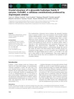
Báo cáo khoa học: Crystal structure of a glycoside hydrolase family 6 enzyme, CcCel6C, a cellulase constitutively produced by Coprinopsis cinerea pot
... The ligand binding cleft of CcCel6C is also wider due to the absence of the bulky tyrosine residue in subsite )3 (Fig 5), and the structures of subsites )1 and )3 of CcCel6C resem- FEBS Journal ... expected to be different from those of known cellobiohydrolases Here, we present the crystal structure of CcCel6C To our knowledge, this is the first report of the crystal structure of a basidiomycete ... FEBS Y Liu et al Structure of C cinerea CcCel6C A R343 D109 D150 R343 D109 D150 B Fig Overall structures of CcCel6C (A) Stereoview of CcCel6C–cellobiose shown as a ribbon model a-Helices, b- strands...
Ngày tải lên: 15/03/2014, 10:20
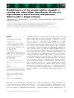
Báo cáo khoa học: Crystal structure of the parasite inhibitor chagasin in complex with papain allows identification of structural requirements for broad reactivity and specificity determinants for target proteases pptx
... inhibitor family Structure 15, 535–543 23 Stubbs MT, Laber B, Bode W, Huber R, Jerala R, ˚ Lenarcic B & Turk V (1990) The refined 2.4 A X-ray crystal structure of recombinant human stefin B in complex ... B) L4 L4 B loop of this enzyme The inhibition of cathepsin B by chagasin is relatively weak, which may be due to the fact that some of the binding energy has to be invested in pushing the occluding ... role of conserved residues of chagasin in the inhibition of cysteine peptidases FEBS Lett 582, 485–490 34 Huang L, Brinen LS & Ellman JA (2003) Crystal structures of reversible ketone-based inhibitors...
Ngày tải lên: 16/03/2014, 04:20