atlat of x ray


handbook of x ray spectrometry revised and expanded by rene van grieken
Ngày tải lên: 10/04/2014, 18:53
Báo cáo hóa học: " Research Article Application of the HLSVD Technique to the Filtering of X-Ray Diffraction Dat" pot
Ngày tải lên: 22/06/2014, 20:20

Báo cáo y học: "Application of Small Angle X-ray Scattering (SAXS) for Differentiation between Normal and Cancerous Breast Tissue"
... using scattered X- rays. J Synchrotron Rad 2000; 7: 348-352. 8. Malden CH, Speller RD, et al. A CdZnTe array for the detection of explosives in baggage by energy dispersive X- ray diffraction ... of their diffraction profiles. 4. Discussion A noticeably shaper peak in the diffraction profile was obtained for adipose compared to other tissues. This is a result of the high levels of ... is expected different peak positions will exist for carcinoma and fibrocystic changes. Our results confirmed this expectation. The peak position of fibrocystic change was close to that of carcinoma....
Ngày tải lên: 02/11/2012, 11:08

Tài liệu Characterization of the Polymorphic Behavior of an Organic Compound Using a Dynamic Thermal and X-ray Powder Diffraction Technique pptx
... melt. Figure 24. DSC of amorphous Form (sample 26) Figure 25. XRPD of amorphous Form (sample 26) Figure 26. DSC of mixture of hydrated Forms I and III (sample 45). Figure 27. DSC of mixture of anhydrous ... 1. Detection of Mixtures of Polymorphs and Potential New Solids Forms. By using the combined and dynamic DSC/ XRPD, several mixtures of 1 disodium salt samples were identified, and the components of these ... apparently formed by refluxing a mixture of Form I and Form IV or by refluxing Form VI in acetone. The proposed interconversions of the various polymorphs of 1 dicarboxylate disodium salt are given in...
Ngày tải lên: 14/02/2014, 03:20

Tài liệu Báo cáo khoa học: X-ray crystallographic and NMR studies of pantothenate synthetase provide insights into the mechanism of homotropic inhibition by pantoate docx
... pantoate-bound form of nPS using X- ray crystal- lography, as described below. Solution and refinement of the structure using X- ray crystallography The crystal structure of the pantoate–nPS complex was solved ... in the vicinity exhibit high flexibility. Preliminary analysis of 15 N relaxation data shows that residues 118–122 have a higher degree of flexibility than other ordered regions of the protein. Unfortunately, ... effects of one of the substrates, i.e. pantoate [8,9]. One of the interesting features of the plant PSs is the presence of a conserved 24 residue insertion in the sequence [9]. This sequence of amino...
Ngày tải lên: 16/02/2014, 09:20

Tài liệu Báo cáo khoa học: Destabilization of psychrotrophic RNase HI in a localized fashion as revealed by mutational and X-ray crystallographic analyses pdf
... to mounting for X- ray diffraction. Structure determination and refinement X- Ray diffraction data sets of the 6·-RNase HI crystal were collected at 100 K using synchrotron radiation at the BL44XU station ... structures of these Ec-RNase HI variants, the position of His124, which corresponds to His126 of So-RNase HI, varies for dif- ferent proteins, because of the intrinsic flexibility of the loop ... mechanism of So-RNase HI A combination of the six thermostabilizing mutations increases the stability of So-RNase HI by 28.8 Cin T m and 27.0 kJặmol )1 in DG(H 2 O). Five of the six substituted...
Ngày tải lên: 18/02/2014, 13:20

Tài liệu Báo cáo khoa học: Molecular determinants of ligand specificity in family 11 carbohydrate binding modules – an NMR, X-ray crystallography and computational chemistry approach doc
... the binding of cellohexaose to CtCBM11. The STD-NMR spectrum of the hexasaccharide in a 20-fold excess over CtCBM11 is shown in Fig. 2 along with the cellohexa- ose reference spectrum. Comparison of both ... in the binding process Fig. 2. STD-NMR of cellohexaose with CtCBM11. (A) Reference 1 H NMR cellohexaose spectrum. (B) STD spectra of the solution of cellohexaose (50 l M) with the protein (5 lM). ... allow determi- nation of the individual contributions of protons aH4a, bH3a, H4b-e and H5b-e to the binding. Fig. 3. Structure of cellohexaose. Relative degrees of saturation of the individual protons...
Ngày tải lên: 18/02/2014, 17:20

Tài liệu Báo cáo khoa học: NMR structure of AII in solution compared with the X-ray structure of AII bound to the mAb Fab131 pptx
... structure of AII in solution with the X- ray structure of AII complexed to the Fab131 mAb. In Fig. 7A, a sequence alignment is represented of the backbone of the conformational ensemble of AII in ... structures of AII to the fragment 3–6 of the X- ray structure of AII. Fig. 6. Structure of a representative folded conformer of AII showing the van der Waals contacts between the side-chains of residues ... yellow the side chains of Val3 and Pro7) (a) with the the X- ray structure of AII in the Fab131–AII complex [30] 3 (b). Ó FEBS 2003 Comparison of the free and bound structure of angiotensin II (Eur....
Ngày tải lên: 20/02/2014, 23:20

Tài liệu Báo cáo Y học: The solution structure and activation of visual arrestin studied by small-angle X-ray scattering pot
... range of 30150 l M (1.36.5 mgặmL )1 ), 90 lL of arrestin solution was mixed with 10 lLof10m M peptide solution to yield a final peptide concentration of 1 m M . For higher concentrations of arrestin ... phosphorylation of the C-terminus of R* and binding by arrestin. Phosphorylation somewhat decreases the ability of R* to signal transducin. Rapid shut- off of R* signalling is then accomplished by binding of arrestin ... Because of the changing oligomeric state of arrestin in these experiments, Guinier analysis was preferred over the use of the distance distribution function, which requires a prior estimation of the...
Ngày tải lên: 22/02/2014, 07:20
Báo cáo khoa học: Determination of thioxylo-oligosaccharide binding to family 11 xylanases using electrospray ionization Fourier transform ion cyclotron resonance mass spectrometry and X-ray crystallography pot
... 4,4 II ,4 III ,4 IV -tetrathio-a-xylopentoside; TRX I, Trichoderma reesei xylanase I; TRX II, Trichoderma reesei xylanase II; Xyl2, b- D-xylobiose; Xyl3, b-D-xylotriose; Xyl4, b-D-xylotetraose; Xyl5, b- D-xylopentaose; Xyl6, ... calorimetry of both domains decreased in the series b-d-xylohexaose (Xyl6) > b-d-xylopentaose (Xyl5) > b-d-xylotetraose (Xyl4) > b-d-xylotriose (Xyl3), with no detectable affinity for b-d-xylobiose ... Param- eters of the measurement and refinement statistics for the CTX–S-Xyl5-Me complex are summarized in Table 4. Atomic co-ordinates for the crystal structures of TRX I, TRX II, CTX, and CTX–S-Xyl5-Me...
Ngày tải lên: 07/03/2014, 17:20

Báo cáo khoa học: The effect of replacing the axial methionine ligand with a lysine residue in cytochrome c-550 from Paracoccus versutus assessed by X-ray crystallography and unfolding ppt
... concentrations of % 5 lm were used. Peroxidase activity was assayed using hydrogen peroxide and guaiacol (O-methoxyphenol, Sigma). The syn- thesis of the fourfold oxidized product of guiacol, 3,3Â-di- methoxy-4,4Â-biphenoquinone ... unfolding of the M100K variant involves the breaking of the Lys-N f -iron bond coupled with the dynamic process of ligand exchange. It thus seems likely that the mechanism of release of the axial ... upon replacing the axial Met ligand with a Lys in the M100K variant of cyt c-550 from P. versutus. We describe three X- ray structures, one of the ferric wild type (wt) and two of the ferric M100K variant....
Ngày tải lên: 07/03/2014, 17:20
Báo cáo khoa học: X-ray crystallography, CD and kinetic studies revealed the essence of the abnormal behaviors of the cytochrome b5 Phe35fiTyr mutant pdf
... Foundation of China. We are grateful to Prof. Li-Wen Niu, Prof. Mai-Kun Teng and Dr Xue-Yong Zhu of the University of Science and Technology of China for their support and help with the X- ray data ... chain conformation of this residue. The shift of the Ca atom of Tyr35 of the Phe35fiTyr mutant from that of Phe35 of the wild-type cyt b 5 is 0.21 A ˚ , within the error limit. The side chain of Tyr35 of the ... structural analysis of bovine cytochrome b 5 at 1.5 A ˚ resolution. Acta Crys- tallogr. D52, 65–76. 23. Xue, L.L. Wang, Y.H. Xie, Y. Yao, P. Wang, W.H. Qian, W. Huang, Z .X. Wu, J. & Xia. Z .X. (1999) Effect of...
Ngày tải lên: 08/03/2014, 10:20
Báo cáo Y học: Determinants of the inhibition of a Taiwan habu venom metalloproteinase by its endogenous inhibitors revealed by X-ray crystallography and synthetic inhibitor analogues pdf
... the Met-turn and active-site consensus HExxHxxGxxH sequence [15–17]. Some organisms and mammalian tissues recently have been reported to contain a number of multidomain proteins, which are related ... stereochemical quality of protein structures. J. Appl. Cryst. 26, 283–291. 43. Bode,W.,Reinemer,P.,Huber,R.,Kleine,T.,Schnierer,S.& Tschesche, H. (1994) The X- ray crystal structure of the catalytic domain of ... Grams,F.,Reinemer,P.,Powers,J.C.,Kleine,T.,Pieper,M., Tschesche, H., Huber, R. & Bode, W. (1995) X- ray structures of human neutrophil collagenase complexed with peptide hydroxamate and peptide thiol inhibitors. Implications for substrate...
Ngày tải lên: 08/03/2014, 23:20
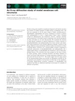
Báo cáo khoa học: An X-ray diffraction study of model membrane raft structures doc
... from a binary mixture of egg-PtdCho and pro- portions of cholesterol of $ 25 mole% (Fig. 5D). The effect of cholesterol on d-spacings of egg-PtdCho bilayers is complex. The presence of only 10 mole% A B C D Fig. ... ternary mixture apparently hinders formation of a gel phase by brainSM in this mixture. Assignment of peak 1 to a structure of pure brainSM can also be excluded on this evidence. The fit of peak ... Laboratory. The X- ray wave- length was 0.154 nm with a beam geometry of $ 0.2 · 0.5 mm in a mica sandwich cell with a surface of 2 · 5 mm and a path length of 0.5 mm. Simultaneous SAXS and WAXS intensities...
Ngày tải lên: 15/03/2014, 23:20
Báo cáo khoa học: X-ray structure of peptidyl-prolyl cis–trans isomerase A from Mycobacterium tuberculosis potx
... rmsd of approxi- mately 1.6 A ˚ from that of Mt PpiA, when the C a atoms are compared using a cut-off of 3.5 A ˚ (with 88% of the Cas matching). This is significantly larger than the r msd of 0.05 ... estimated k cat /K m of 2.0 Ã 10 6 M )1 ặs )1 .The X- ray structure of PpiA was solved by molecular replace- ment, and r efined t o a resoluti on of 2.6 A ˚ with R and R free values of 21.3% and 22.9%, ... the Correspondence to S. Mowbray, Department of Molecular Biology, Swedish University of Agricultural Sciences, Uppsala Biomedical Center, Box 590, SE-751 24 Uppsala, Sweden. Fax: +46 18 53 69 71, Tel.:...
Ngày tải lên: 16/03/2014, 18:20

Báo cáo Y học: The role of zinc in the methylation of the coenzyme M thiol group in methanol:coenzyme M methyltransferase from Methanosarcina barkeri New insights from X-ray absorption spectroscopy doc
... zinc. XANES spectra of wild-type and mutants As shown in the following, the interpretation of the EXAFS of the mutant enzymes is confirmed by a comparative analysis of the zinc K-edge spectra (XANES ... Mta, methanol:coenzyme M methyltransferase; MtaA, protein subunit of Mta; XANES, X- ray absorption near edge structure; XAS, X- ray absorption spectroscopy. (Received November 2001, revised 15 February ... curve-fitting of unfiltered k space the in-house software SIMX was used. By curve-fitting of various EXAFS spectra we found consistently that DE 0 refined to a value of % 9665 eV; this value has been fixed...
Ngày tải lên: 17/03/2014, 23:20
Báo cáo khoa học: X-ray structure of glucose/galactose receptor from Salmonella typhimurium in complex with the physiological ligand, (2R)-glyceryl-b-D-galactopyranoside pdf
... 4990 E-mail: mowbray@xray.bmc.uu.se Website: http://xray.bmc.uu.se/ (Received 13 December 2008, revised 31 January 2009, accepted 2 February 2009) doi:10.1111/j.1742-4658.2009.06945 .x Periplasmic ... Structure of the periplasmic glucose ⁄ galactose receptor of Salmonella typhimurium . Receptor 1 , 41–53. 21 Zou JY, Flocco MM & Mowbray SL (1993) The 1.7 A ˚ refined X- ray structure of the periplasmic ... binding sites of the Escherichia coli galactose chemoreceptor protein. Science 242, 1290– 1295. 25 Flocco MM & Mowbray SL (1994) The 1.9 A ˚ X- ray structure of a closed unliganded form of the periplasmic glucose...
Ngày tải lên: 23/03/2014, 04:21