amp 947 scintigraphy and magnetic resonance

Pocket Atlas of Sectional Anatomy Computed Tomography and Magnetic Resonance Imaging - Volume II Thorax, Heart, Abdomen, and Pelvis (Part 1 ) pptx
... III Pocket Atlas of Sectional Anatomy Computed Tomography and Magnetic Resonance Imaging Volume II Thorax, Heart, Abdomen, and Pelvis Torsten B Moeller, MD Department of Radiology Caritas ... processing and storage 12345 Moeller, Sectional Anatomy © 2007 Thieme All rights reserved Usage subject to terms and conditions of license V Dedicated to our parents, Alfred and Friedel Möller and ... in MRI and CT visualization of the vascular system have also further improved For example, stepwise shifts of the table top have become standard procedures in angiography of the pelvis and leg...
Ngày tải lên: 29/06/2014, 09:21
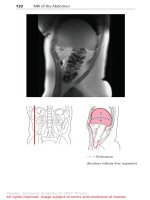
Pocket Atlas of Sectional Anatomy Computed Tomography and Magnetic Resonance Imaging - Volume II Thorax, Heart, Abdomen, and Pelvis (Part 2 ) doc
... 31 32 Rib Intercostal artery, vein and nerve Intercostalis muscles Costodiaphragmatic recess Latissimus dorsi muscle Adrenal gland Right kidney Renal artery and vein Ureter Quadratus lumborum ... 22 Diaphragm (lumbar part) 23 Splenic artery and vein 24 Adrenal gland 25 Left kidney 26 Intertransversarii muscles (lateral lumbar) 27 Renal artery and vein 28 Latissimus dorsi muscle 29 Erector ... subject to terms and conditions of license 165 166 MR Angiography of the Splenic and Portal Veins Moeller, Sectional Anatomy © 2007 Thieme All rights reserved Usage subject to terms and conditions...
Ngày tải lên: 29/06/2014, 09:21
Báo cáo y học: "Correlation of histopathological findings and magnetic resonance imaging in the spine of patients with ankylosing spondylitis" potx
... Correlation of histopathological findings and magnetic resonance imaging Patient no HLA-B27 Edema (%) Cellular infiltration Microvessel density Edema detected by magnetic resonance imaging Zygapophyseal ... Positive correlation of bone marrow edema in histopathological assessment and magnetic resonance imaging (a) Magnetic resonance imaging (T2 imaging sequence) of a zygapophyseal joint with bone ... Negative correlation of bone marrow edema in histopathological assessment and magnetic resonance imaging (a) Magnetic resonance imaging of imaging zygapophyseal joints (cyan arrows) without detectable...
Ngày tải lên: 09/08/2014, 08:22

báo cáo khoa học: " N-hexanoyl chitosan stabilized magnetic nanoparticles: Implication for cellular labeling and magnetic resonance imaging" pps
... size and morphology of IOPs and MC-IOPs were observed by TEM (JEM-1230, JEOL, Japan) and HRTEM (QUANTA 200F, FEI, USA) The sample for TEM analysis was obtained by placing a drop of IOPs and MC-IOPs ... (curve c), and MCIOPs (curve d) IOPs exhibit strong bands in the low frequency region below 800 cm-1 due to the oxide skeleton The characteristic bands of modified chitosan, amide I, II, and III ... spectra of IOPs have weak bands The spectrum is consistent with magnetic (Fe3O4) and the signals associated to the magnetite appear as broad features at 408.9, 571.5 and 584.5 cm-1 [4] Figure...
Ngày tải lên: 11/08/2014, 00:22

Báo cáo y học: "Soft-tissue perineurioma of the retroperitoneum in a 63-year-old man, computed tomography and magnetic resonance imaging findings: a case report" doc
... contributions MY conceived the study YK and RM performed the literature review AA and KK performed histopathologic and immunohistochemical analyses All authors read and approved the final version of ... 22:227-231 doi:10.1186/1752- 1947- 4-290 Cite this article as: Yasumoto et al.: Soft-tissue perineurioma of the retroperitoneum in a 63-year-old man, computed tomography and magnetic resonance imaging findings: ... soft tissue, intramural, sclerosing, and reticular Soft-tissue perineuriomas are the most common subtype and show distinctive morphologic, ultrastructual, and immunophenotypic features that distinguish...
Ngày tải lên: 11/08/2014, 03:21

Báo cáo y học: "Soft-tissue perineurioma of the retroperitoneum in a 63-year-old man, computed tomography and magnetic resonance imaging findings: a case report." pps
... contributions MY conceived the study YK and RM performed the literature review AA and KK performed histopathologic and immunohistochemical analyses All authors read and approved the final version of ... 22:227-231 doi:10.1186/1752- 1947- 4-290 Cite this article as: Yasumoto et al.: Soft-tissue perineurioma of the retroperitoneum in a 63-year-old man, computed tomography and magnetic resonance imaging findings: ... soft tissue, intramural, sclerosing, and reticular Soft-tissue perineuriomas are the most common subtype and show distinctive morphologic, ultrastructual, and immunophenotypic features that distinguish...
Ngày tải lên: 11/08/2014, 07:20

Báo cáo y học: " Change in CD3 positive T-cell expression in psoriatic arthritis synovium correlates with change in DAS28 and magnetic resonance imaging synovitis scores following initiation of biologic therapy-a single centre, open-label study" doc
... were measured at weeks and 12, including 28 and 68 tender (TJC) and 28 and 66 swollen joint counts (SJC), patient pain and disease to 100 mm visual analogue scale (VAS) and the Health Assessment ... in a DAS28 moderate responder (Patient 1) and non-responder (Patient 2) A and B are baseline and Week 12 images of Patient 1, and C and D are baseline and Week 12 images of Patient respectively ... Patients were 50% and 66.6% female, had a mean age (range) of 43.2 (27 to 60) and 48.7 (26 to 64) years and a mean disease duration of 9.1 (1 to 42) and 7.5 (1 to 29) years in the anakinra and etanercept...
Ngày tải lên: 12/08/2014, 15:22
The application of electromagnetic theory in microwave therapy and magnetic resonance imaging
... Denier, who, in 1936, employed combined L-band microwave (80 cm) and X-rays [11] Subsequently, Brunner-Ornzsteini and Randa reported that the use of L-band microwave (60 cm) combined with X-rays ... microwave cancer therapy and magnetic resonance imaging, followed by thesis organization and publications In Chapter 2, an invasive microwave breast cancer therapy is proposed, and good heating effect ... China, Jul 28-30, 2010 Dandan Liang, Hon Tat Hui, and Tat Soon Yeo, “Study on the Decoupling of Stacked Phased Array Coils for Magnetic Resonance Imaging,” Progress In Electromagnetics Research Symposium,...
Ngày tải lên: 09/09/2015, 10:20

Tài liệu Magnetic resonance imaging and gynecological devices doc
... Lack of interaction between magnetic resonance imaging and the copper-T380A IUD Contraception 1997;55:169–73 543 [13] Zieman M, Kanal E Copper T 380A IUD and magnetic resonance imaging Contraception ... superparamagnetic or ferromagnetic) In the presence of discordant (antiparallel) magnetization, the substances are classified as exhibiting negative magnetic susceptibility and are called diamagnetic ... or implants Material and methods The authors researched in PubMed-Medline/Ovid using the following keywords: Magnetic Resonance Imaging, Intrauterine Devices, Implanon® and Essure® This article...
Ngày tải lên: 13/02/2014, 07:20

báo cáo hóa học:" Dynamic magnetic resonance imaging in assessing lung function in adolescent idiopathic scoliosis: a pilot study of comparison before and after posterior spinal fusion" doc
... apex and counter pressures at opposite ends under SSEP monitoring Rod estimation and preliminary contouring was then made Decortication of facet joints and transverse processes was made and bone ... into axial and coronal planes so that motions of the chest wall and diaphragm could be assessed The chest wall and diaphragmatic motions were measured in antero-posterior, left-right and cranio-caudal ... the highest point of the diaphragm and a line parallel to the lung apex (Fig 3a and 3b) All the lung volume, chest wall and diaphragmatic dimensions in the right and left hemithorax were measured...
Ngày tải lên: 20/06/2014, 01:20

Báo cáo y học: "Autologous chondrocyte implantation for cartilage repair: monitoring its success by magnetic resonance imaging and histology" ppt
... O’Driscoll; MRI, magnetic resonance imaging; n/a not applicable; N/A not available with standard and polarised light and images captured and digitised using a closed-circuit television and Image Grabber ... in hyaline-like cartilage (a) (sample 22) and (b) (H) (sample 2), whereas in specimens with a more fibrocartilaginous morphology (b) (F) (sample 2) and (c) (sample 15), it was predominantly homogeneous ... whom biopsy specimens were obtained and their histology and MRI scores Patient and sample no Patient’s age at ACI (years) 20 20 25 28 Sex Interval between graft and biopsy (months) M Treatment Location...
Ngày tải lên: 09/08/2014, 01:21
Báo cáo y học: "Magnetic resonance imaging changes of sacroiliac joints in patients with recent-onset inflammatory back pain: inter-reader reliability and prevalence of abnormalities" potx
... changes and structural changes and the different localization of these changes Pathological changes of interest were defined as inflammation and structural changes including erosions, sclerosis and ... capsule, the joint space and the retro-auricular ligaments Firstly, in different sessions, MRI scans were reviewed and scored together by two observers (LHD and RW) and discrepancies in scoring ... all sequences) Inflammation and sclerosis were scored on the iliac and sacral side of both SI joints separately Erosions and ankylosis were scored for the entire left and right SI joints Active...
Ngày tải lên: 09/08/2014, 07:20

Báo cáo y học: "Long term evaluation of disease progression through the quantitative magnetic resonance imaging of symptomatic knee osteoarthritis patients: correlation with clinical symptoms and radiographic change" pps
... demographic, clinical, radiological, and biomarker features, and presented as mean ± standard deviation Non-parametric Wilcoxon one-sample tests, one-sample and two-sample Student t tests, chi-squares, ... Early morning fasting second void urine samples were collected at baseline, month 6, 12 and 24 (exit) The samples were taken between hours and 21 hours and not necessarily collected at the same ... JPR, JMP, JMM, JFB, GAC, and JPP analyzed and interpreted the data JPR, JMP and JPP prepared the manuscript JPR and GAC performed the statistical analysis All authors read and approved the final...
Ngày tải lên: 09/08/2014, 07:20

Báo cáo y học: "proximal interphalangeal joints in rheumatoid arthritis: a comparison with magnetic resonance imaging, conventional radiography and clinical examination" pdf
... T1-weighted magnetic resonance images were obtained (b) before and (c) after contrast administration (grade synovitis) MRI, magnetic resonance imaging; RA, rheumatoid arthritis second destruction and ... fifth MCP joints and the second to fifth PIP joints of the dominant hand: the dorsal, radial and palmar aspects of the second MCP joint; the dorsal and palmar aspects of the third and fourth MCP ... longitudinal and (b) the transverse planes shows both signs of destruction (grade 2) and inflammation (grade 3) Axial T1weighted magnetic resonance images were obtained (c) before and (d) after...
Ngày tải lên: 09/08/2014, 07:20

Báo cáo y học: "Are bone erosions detected by magnetic resonance imaging and ultrasonography true erosions? A comparison with computed tomography in rheumatoid arthritis metacarpophalangeal joints" pptx
... echo sequence were done in the axial and coronal planes with a slice thickness of 0.4 mm, and these were used for image evaluation (Figures and 2) Magnetic resonance images were evaluated by a ... in evaluating magnetic resonance images of RA finger joints Statistical analysis With CT as the standard reference method, the sensitivity, specificity and accuracy of MRI, US and radiography ... overall sensitivity, specificity and accuracy were 19%, 100% and 81%, respectively For MRI, the corresponding values were 68%, 96% and 89%, and for US they were 42%, 91% and 80% (Table 2) To evaluate...
Ngày tải lên: 09/08/2014, 08:22

Báo cáo y học: "The association between patellar alignment on magnetic resonance imaging and radiographic manifestations of knee osteoarthritis" ppsx
... femoral sulcus and through the patella, and measured the distance between the lateral border of the patella and this vertical line (a) and between the medial border of the patella and this vertical ... pixels; and coronal and axial spinecho fat-suppressed proton density-weighted and T2weighted images (TR 2,200 ms; TE 20/80 ms) with a slice thickness of mm, a mm interslice gap, one excitation, and ... by magnetic resonance imaging (MRI) [19-21] Muellner and colleagues [19] performed measurements analogous to those used in Xray evaluation with MRI images obtained with knees flexed to 20° and...
Ngày tải lên: 09/08/2014, 10:20

Báo cáo y học: "Cartilage markers and their association with cartilage loss on magnetic resonance imaging in knee osteoarthritis: the Boston Osteoarthritis Knee Study" doc
... excitation, and the same FOV and matrix Cartilage on MRI was scored paired and unblinded to sequence on 14 plates (anterior, central, and posterior femur; anterior, central, and posterior tibia; and ... its design and coordination, and drafted the manuscript JL and ML carried out the statistical analyses JD carried out the assays AG and DG read and interpreted the MRIs DB, MN, RP, DE, and DF participated ... which the original WORMS score of and were collapsed to 0, the original scores of and were collapsed to 1, and the original scores of 4, 5, and were considered 2, 3, and 4, respectively Cartilage...
Ngày tải lên: 09/08/2014, 10:21
Báo cáo y học: "Ultrasonography, magnetic resonance imaging, radiography, and clinical assessment of inflammatory and destructive changes in fingers and toes of patients with psoriatic arthritis" pot
... hands and feet in a posterior-anterior projection was performed within a month of the US X-rays of both hands and feet (2nd–5th MCP, PIP, DIP, and MTP joints) were assessed for bone erosions and ... x-ray and not by US were in the non-MRIexamined hand However, x-ray erosions (all in PsA: MCP and DIP joints) and proliferations (both PsA: DIP joints) were located in the MRI-examined hand and ... ultrasonography (US) and magnetic resonance imaging (MRI) appear promising MRI can detect inflammation and bone destruction in joints earlier than projection radiography (x-ray) in PsA, RA, and spondyloarthritis...
Ngày tải lên: 09/08/2014, 10:21
Báo cáo y học: "Detection of bone erosions in rheumatoid arthritis wrist joints with magnetic resonance imaging, computed tomography and radiography" ppt
... tomography and (c, d) T1patient weighted magnetic resonance imaging in the (a, c) coronal and (b, d) axial planes A bone erosion at the distal radius is seen on both computed tomography and magnetic resonance ... and intermodality agreements of single and total erosion volume, measured on computed tomography (CT) and magnetic resonance imaging (MRI) Reading A (mm3) Reading B (mm3) Mean of readings A and ... Computerized measurement of magnetic resonance imaging erosion volumes in patients with rheumatoid arthritis: a comparison with existing magnetic resonance imaging scoring systems and standard clinical outcome...
Ngày tải lên: 09/08/2014, 10:23
