Atlas of the Diabetic Foot - part 10 pps

Atlas of the Diabetic Foot - part 10 pps
... 1 ANATOMY OF THE FOOT Atlas of the Diabetic Foot. N. Katsilambros, E. Dounis, P. Tsapogas and N. Tentolouris Copyright © 2003 John Wiley & Sons, Ltd. ISBN: 0-4 7 1-4 867 3-6 210 Atlas of the Diabetic ... 35 Anatomy of the Foot 215 Figure A1 Dorsal aspect of the bones in the foot Figure A3 Plain radiograph of the foot shown in lateral...
Ngày tải lên: 10/08/2014, 18:21
Atlas of the Diabetic Foot - part 2 ppsx
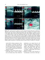
... ide of a three-layer cus- tom-made insole used to of oad pressure on the forefoot. The upper layer is composed of cross-linked polyethylene foam, the mid- dle layer of polyurethane, and the lower ... overleaf ) 32 Atlas of the Diabetic Foot should fully understand the risks posed by the loss of protective sensation or an inad- equate blood supply to their feet....
Ngày tải lên: 10/08/2014, 18:21

Atlas of the Diabetic Foot - part 6 ppsx
... base on the medial aspect of the right hallux Figure 6.19 Osteomyelitis of the condyle in the proximal phalanx of the hallux of the foot shown in Figure 6.18 128 Atlas of the Diabetic Foot Figure ... Heel ulcer in the patient whose foot is shown in Figure 6.14. The yellowish appearance of the bed of the ulcer is indicative of ischemia 118 Atlas...
Ngày tải lên: 10/08/2014, 18:21

Atlas of the Diabetic Foot - part 1 pot
... in the field of diabetes and the diabetic foot. Atlas of the Diabetic Foot Professor Nicholas Katsilambros, MD Director of the 1 st Department of Propaedeutic Medicine and the Diabetic Centre Athens ... VASCULAR DISEASE ASSESSMENT OF THE VASCULAR STATUS IN PATIENTS WITH DIABETES The prevalence of peripheral vascular dis- ease in diabetic patients is 15–30...
Ngày tải lên: 10/08/2014, 18:21

Atlas of the Diabetic Foot - part 3 pdf
... revealed osteomyeli- tis of the first metatarsal head extend- ing to the base of the proximal pha- lanx of the great toe. Cultures of the base of the ulcer revealed the presence of Staphylococcus ... causing sublux- ation of the phalangeal bases. Contractures of tendons and joint capsules result in fixa- tion of the deformity. Due to the deformity of the...
Ngày tải lên: 10/08/2014, 18:21
Atlas of the Diabetic Foot - part 4 doc
... 93% of all foot ulcers. Almost 20% of the ulcers developed under the hallux, 22% over the metatarsal heads, 26% on the tips of the toes and 16% on the dorsum of the toes. Ulcer under the hal- lux ... radio- graph excluded osteomyelitis. Therapeu- tic footwear was prescribed and the ulcer healed in 6 weeks. The forefoot is the usual site for ulcer- ation. In one...
Ngày tải lên: 10/08/2014, 18:21

Atlas of the Diabetic Foot - part 5 potx
... metatarso- phalangeal joint of left hallux of the patient whose foot is shown in Figures 6.4 and 6.5 weak and the ankle brachial index was 0.6. At the base of the ulcer the fascia of the dorsum of the ... significant ankle edema. The ulcer was a result of fric- tion between the foot and the forepart of the patient’s narrow shoe upper (vamp) follow- ing...
Ngày tải lên: 10/08/2014, 18:21

Atlas of the Diabetic Foot - part 7 ppt
... mid-tarsal disar- ticulation EXTENSIVE WET GANGRENE OF THE FOOT A 51-year-old male patient with type 1 dia- betes diagnosed at the age of 25 years was admitted to the Vascular Surgery Depart- ment ... GANGRENE OF THE TOES A 54-year-old male patient with type 2 dia- betes diagnosed at the age of 49 years was admitted to the Vascular Surgery Depart- ment because of wet g...
Ngày tải lên: 10/08/2014, 18:21

Atlas of the Diabetic Foot - part 8 ppt
... sed- imentation rate (74 mm/h) and mild leuko- cytosis were found, therefore the possi- bility of osteomyelitis was high. A mag- netic resonance imaging-T1-weighted sagit- tal image of the foot ... initially, and the continua- tion of oral treatment for a prolonged period (at least 6 weeks). 166 Atlas of the Diabetic Foot Figure 8.16 Left neuro-osteoarthropathic foot...
Ngày tải lên: 10/08/2014, 18:21

Atlas of the Diabetic Foot - part 9 pptx
... instability and development of varus deformity of the foot. 204 Atlas of the Diabetic Foot Figure 9.21 Plain radiograph of chronic neuro-osteoarthropathy in the patient whose foot is illustrated in ... lateral exostosis of the proximal phalanx of the second toe are all evident 200 Atlas of the Diabetic Foot fragments in the talonavicular joint dor-...
Ngày tải lên: 10/08/2014, 18:21