Atlas of the Diabetic Foot - part 9 pptx

Atlas of the Diabetic Foot - part 9 pptx
... development of varus deformity of the foot. 204 Atlas of the Diabetic Foot Figure 9. 21 Plain radiograph of chronic neuro-osteoarthropathy in the patient whose foot is illustrated in Figures 9. 19 and 9. 20. ... seen 198 Atlas of the Diabetic Foot Figure 9. 13 Postoperative radiographs of the condition shown in Figure 9. 12; fusion of the tarso...
Ngày tải lên: 10/08/2014, 18:21

Atlas of the Diabetic Foot - part 1 pot
... in the field of diabetes and the diabetic foot. Atlas of the Diabetic Foot Professor Nicholas Katsilambros, MD Director of the 1 st Department of Propaedeutic Medicine and the Diabetic Centre Athens ... VASCULAR DISEASE ASSESSMENT OF THE VASCULAR STATUS IN PATIENTS WITH DIABETES The prevalence of peripheral vascular dis- ease in diabetic patients is 15–30...
Ngày tải lên: 10/08/2014, 18:21
Atlas of the Diabetic Foot - part 2 ppsx
... ide of a three-layer cus- tom-made insole used to of oad pressure on the forefoot. The upper layer is composed of cross-linked polyethylene foam, the mid- dle layer of polyurethane, and the lower ... Amsterdam, The Nether- lands, 199 9. 2. Boulton AJM, Greis FA, Jervell JA. Guide- lines for the diagnosis and outpatient man- agement of diabetic peripheral neuropathy. Dia...
Ngày tải lên: 10/08/2014, 18:21

Atlas of the Diabetic Foot - part 3 pdf
... revealed osteomyeli- tis of the first metatarsal head extend- ing to the base of the proximal pha- lanx of the great toe. Cultures of the base of the ulcer revealed the presence of Staphylococcus ... causing sublux- ation of the phalangeal bases. Contractures of tendons and joint capsules result in fixa- tion of the deformity. Due to the deformity of the...
Ngày tải lên: 10/08/2014, 18:21
Atlas of the Diabetic Foot - part 4 doc
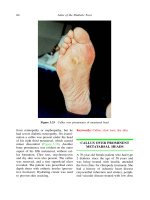
... 93 % of all foot ulcers. Almost 20% of the ulcers developed under the hallux, 22% over the metatarsal heads, 26% on the tips of the toes and 16% on the dorsum of the toes. Ulcer under the hal- lux ... radio- graph excluded osteomyelitis. Therapeu- tic footwear was prescribed and the ulcer healed in 6 weeks. The forefoot is the usual site for ulcer- ation. In o...
Ngày tải lên: 10/08/2014, 18:21

Atlas of the Diabetic Foot - part 5 potx
... metatarso- phalangeal joint of left hallux of the patient whose foot is shown in Figures 6.4 and 6.5 weak and the ankle brachial index was 0.6. At the base of the ulcer the fascia of the dorsum of the ... significant ankle edema. The ulcer was a result of fric- tion between the foot and the forepart of the patient’s narrow shoe upper (vamp) follow- ing...
Ngày tải lên: 10/08/2014, 18:21

Atlas of the Diabetic Foot - part 6 ppsx
... base on the medial aspect of the right hallux Figure 6. 19 Osteomyelitis of the condyle in the proximal phalanx of the hallux of the foot shown in Figure 6.18 128 Atlas of the Diabetic Foot Figure ... Heel ulcer in the patient whose foot is shown in Figure 6.14. The yellowish appearance of the bed of the ulcer is indicative of ischemia 118 Atl...
Ngày tải lên: 10/08/2014, 18:21

Atlas of the Diabetic Foot - part 7 ppt
... GANGRENE OF THE TOES A 54-year-old male patient with type 2 dia- betes diagnosed at the age of 49 years was admitted to the Vascular Surgery Depart- ment because of wet gangrene involving the toes of ... mid-tarsal disar- ticulation EXTENSIVE WET GANGRENE OF THE FOOT A 51-year-old male patient with type 1 dia- betes diagnosed at the age of 25 years was admitted to...
Ngày tải lên: 10/08/2014, 18:21

Atlas of the Diabetic Foot - part 8 ppt
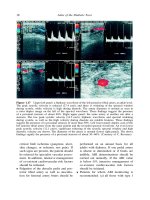
... sed- imentation rate (74 mm/h) and mild leuko- cytosis were found, therefore the possi- bility of osteomyelitis was high. A mag- netic resonance imaging-T1-weighted sagit- tal image of the foot ... technetium -9 9 m ( 99 Tc) phosphonate scan. Images obtained dur- ing the flow phase are shown in the left upper panel of Figure 8.37; during this phase a series of 3-s image acq...
Ngày tải lên: 10/08/2014, 18:21

Atlas of the Diabetic Foot - part 10 pps
... ISBN: 0-4 7 1-4 867 3-6 210 Atlas of the Diabetic Foot Figure 9. 32 Plain postoperative radiograph of the right foot of the patient whose feet are illustrated in Figures 9. 29 9. 31. Arthrodesis of the ... The Charcot Foot 2 09 Figure 9. 30 Lateral view of Figure 9. 29 Figure 9. 31 Plain radiograph of neuro-osteoarthropathy of the right foot...
Ngày tải lên: 10/08/2014, 18:21
- cracking the toefl ibt part 9
- the turn of the screw 1999 part 1
- planning atlas of the lower mekong river basin
- social atlas of the lower mekong basin
- the turn of the screw summary chapter 9
- lord of the flies chapter 6 9 summary
- giáo trình công nghệ lọc dầu part 9 pptx 162
- the lord of the rings pdf part 1
- master of the world 1961 part 1