Atlas of the Diabetic Foot - part 2 ppsx
Atlas of the Diabetic Foot - part 2 ppsx
... of Foot Ulcers 33 Figure 2. 11 Upper s ide of a three-layer cus- tom-made insole used to of oad pressure on the forefoot. The upper layer is composed of cross-linked polyethylene foam, the mid- dle ... overleaf ) 32 Atlas of the Diabetic Foot should fully understand the risks posed by the loss of protective sensation or an inad- equate blood supply to their f...
Ngày tải lên: 10/08/2014, 18:21

Atlas of the Diabetic Foot - part 6 ppsx
... base on the medial aspect of the right hallux Figure 6.19 Osteomyelitis of the condyle in the proximal phalanx of the hallux of the foot shown in Figure 6.18 128 Atlas of the Diabetic Foot Figure ... Sons, Ltd. ISBN: 0-4 7 1-4 867 3-6 120 Atlas of the Diabetic Foot Figure 6 .21 Neuro-ischemic ulcers on the dorsum of claw toes Figure 6 .2...
Ngày tải lên: 10/08/2014, 18:21

Atlas of the Diabetic Foot - part 1 pot
... in the field of diabetes and the diabetic foot. Atlas of the Diabetic Foot Professor Nicholas Katsilambros, MD Director of the 1 st Department of Propaedeutic Medicine and the Diabetic Centre Athens ... 21 3 Appendix 2 Manufacturers of Preventive and Therapeutic Footwear 21 7 Index 22 1 12 Atlas of the Diabetic Foot Figure 1.10 Qualitative analys...
Ngày tải lên: 10/08/2014, 18:21
Atlas of the Diabetic Foot - part 3 pdf
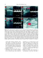
... revealed osteomyeli- tis of the first metatarsal head extend- ing to the base of the proximal pha- lanx of the great toe. Cultures of the base of the ulcer revealed the presence of Staphylococcus ... AMPUTATION A 7 2- year-old male patient with type 2 dia- betes diagnosed at the age of 56 years and Figure 3.30 X-ray image of the foot illustrated in Figures...
Ngày tải lên: 10/08/2014, 18:21
Atlas of the Diabetic Foot - part 4 doc
... 93% of all foot ulcers. Almost 20 % of the ulcers developed under the hallux, 22 % over the metatarsal heads, 26 % on the tips of the toes and 16% on the dorsum of the toes. Ulcer under the hal- lux ... lipoidica 88 Atlas of the Diabetic Foot Figure 5 .2 Therapeutic footwear prescribed for the patient whose foot is shown in Figure 5.1. Among the m...
Ngày tải lên: 10/08/2014, 18:21

Atlas of the Diabetic Foot - part 5 potx
... Diabetic Foot Figure 5 .23 Plain radiograph of the right foot of the patient whose foot is shown in Figure 5 .21 . Osteomyelitis of the fifth metatarsal head and the proxi- mal phalanx of the fifth ... & Sons, Ltd. ISBN: 0-4 7 1-4 867 3-6 104 Atlas of the Diabetic Foot Figure 5.30 Healed ulcer of the p atient whose feet are shown in Figures 5...
Ngày tải lên: 10/08/2014, 18:21

Atlas of the Diabetic Foot - part 7 ppt
... 16,000/l. Figure 7 .23 Wet gangrene of the right foot. Redness and edema due to infection extends as far as the lower third of the tibia. (Courtesy of E. Bastounis) 146 Atlas of the Diabetic Foot Figure 7 .26 ... GANGRENE OF THE TOES A 54-year-old male patient with type 2 dia- betes diagnosed at the age of 49 years was admitted to the Vascular Surgery Depar...
Ngày tải lên: 10/08/2014, 18:21

Atlas of the Diabetic Foot - part 8 ppt
... for 2 weeks initially, and the continua- tion of oral treatment for a prolonged period (at least 6 weeks). 166 Atlas of the Diabetic Foot Figure 8.16 Left neuro-osteoarthropathic foot of the ... sed- imentation rate (74 mm/h) and mild leuko- cytosis were found, therefore the possi- bility of osteomyelitis was high. A mag- netic resonance imaging-T1-weighted sagit- ta...
Ngày tải lên: 10/08/2014, 18:21

Atlas of the Diabetic Foot - part 9 pptx
... development of varus deformity of the foot. 20 4 Atlas of the Diabetic Foot Figure 9 .21 Plain radiograph of chronic neuro-osteoarthropathy in the patient whose foot is illustrated in Figures 9.19 and 9 .20 . ... to of oad the pressure from the ulcerated area and to accommodate the deformity. The ulcer healed in 12 weeks (Figure 9 .23 ). Within the next 2...
Ngày tải lên: 10/08/2014, 18:21

Atlas of the Diabetic Foot - part 10 pps
... formation talonavicular joint Chopart dislocation 69 collapse 20 1, 20 2 destruction 20 3, 20 4 fragments 199, 20 0 neuro-osteoarthropathy 20 3 talus resorption 20 6, 20 7, 20 8, 20 9, 21 0, 21 1, 21 2 tardus pardus waveform ... 20 8, 21 0 bone fragments 20 8, 20 9 disarticulation 181 edema 53, 110, 1 12 deep-tissue infection 1 62, 163 neuro-osteoarthropathy 20 4, 20 5, 2...
Ngày tải lên: 10/08/2014, 18:21
- lord of the rings pdf part 2
- the art of negotiation in diplomacy part 2
- under the hood of net memory management part 2 pdf
- the other side of love nigerian movie part 2
- lord of the rings war in the north walkthrough part 2
- finest hour the battle of britain 2000 u2014 part 2
- the end of time doctor who part 2 watch online
- ace the toefl essay part 2