Manual of Diagnostic Ultrasound in Infectious Tropical Diseases - part 2 pptx

Manual of Diagnostic Ultrasound in Infectious Tropical Diseases - part 2 pptx
... disadvantage of dissolving rubber or plastic parts of the transducer. 22 2 Typical Sonographic Findings in Inflammatory Diseases – emigration of leukocytes, neutrophils, or monocytes (= cell ular in ltra- tion) Abscesses ... 2. 2 Ultrasonic Findings 23 Fig. 2. 1. Acute colitis. Segment of the descending colon. Power Doppler shows multiple color pixels in the wall, indicat...
Ngày tải lên: 10/08/2014, 16:22

Manual of Diagnostic Ultrasound in Infectious Tropical Diseases - part 5 pptx
... pathol- ogy, but may indicate the presence of a non-neoplastic gastrointestinal involvement. In infectious diarrhea, ultrasound shows five sonographic patterns in 80% of cases: 1. Dilated intestinal ... C virus infection. With a high incidence in southeast Asia and parts of Africa, HCC is one of the most common malignant tumors worldwide. 3 .2. 2.3 Ultrasound Findings In...
Ngày tải lên: 10/08/2014, 16:22

Manual of Diagnostic Ultrasound in Infectious Tropical Diseases - part 1 pps
... UltrasonicFindings 22 2. 3 Organ-relatedUltrasonicFindings 27 2. 3.1 LymphNodes 27 2. 3 .2 Spleen 32 2.3.3 LungandPleura 36 2. 3.4 LiverandBiliaryTract 42 2.3.5 GastrointestinalTract 49 2. 3.6 Kidney ... 159 4 .2 UltrasoundinBrainInfectioninNeonatesandInfants 161 4 .2. 1 Cytomegalovirus 1 62 4 .2. 2 Toxoplasmosis 1 62 4 .2. 3 HerpesSimplexVirus 1 62 4 .2. 4 CongenitalRubella 16...
Ngày tải lên: 10/08/2014, 16:22

Manual of Diagnostic Ultrasound in Infectious Tropical Diseases - part 3 doc
... anterior mediastinum is scanned on both sides of the sternum. 34 2 Typical Sonographic Findings in Inflammatory Diseases Table 2. 2. Major infectious (tropical) diseases affecting the spleen Tub ... pleura. 2. 3 Organ-related Ultrasonic Findings 29 2. 3.1.4 Pathologic Findings Lymph nodes are nearly always involved in inflammatory diseases, either directly by the in fectiou...
Ngày tải lên: 10/08/2014, 16:22

Manual of Diagnostic Ultrasound in Infectious Tropical Diseases - part 4 pps
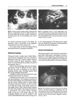
... exclusion of such a lesion is not possible (Figs. 2. 46 2. 51). 60 2 Typical Sonographic Findings in Inflammatory Diseases Fig. 2. 62. Chronic pyelonephritis. Cir- rhotic kidney with a thin inhomoge- neous ... Sonographic Findings in Inflammatory Diseases Fig. 2. 55. In ammatory tumor. Con- glomerate of in amed mesentery, in- volved sections of the bowel, fluid, and short...
Ngày tải lên: 10/08/2014, 16:22

Manual of Diagnostic Ultrasound in Infectious Tropical Diseases - part 6 ppt
... majority of patients overcoming either the classic or the hemor- rhagic form of the disease remain in a considerably weak state for a period of several weeks. 3 .2. 3.4 Laboratory Findings Findings include ... which is common in the tropical and subtropical areas throughout the world, having its maximumincidence at the end of the rainy season. A significant increase in the inci...
Ngày tải lên: 10/08/2014, 16:22

Manual of Diagnostic Ultrasound in Infectious Tropical Diseases - part 8 docx
... accompanying portal veins. 122 3 Ultrasound Diagnosis of Special Infectious and Parasitic Diseases Fig. 3.57. Brazilian25-year-old male. Ul- trasound of scrotal contents in B- mode with 3.5-MHz probe ... 1 32 3 Ultrasound Diagnosis of Special Infectious and Parasitic Diseases Fig. 3.61. Macrosc opic examination of the liver, show ing intense fibrous peri- portal thicken...
Ngày tải lên: 10/08/2014, 16:22

Manual of Diagnostic Ultrasound in Infectious Tropical Diseases - part 9 pdf
... in neonates and infants can be clinically silent. Once clinical suspicion is aroused and the physical examination and laboratory tests are in keeping with an infectious process, plain film ra- diographs ... of hyperosmolar saline so- lution. c after injection. d after reaspiration 1 52 3 Ultrasound Diagnosis of Special Infectious and Parasitic Diseases Fig. 3.87. Pancreatic hy...
Ngày tải lên: 10/08/2014, 16:22
General ultrasound In the critically ill - part 2 pdf
... Conducting an Ultrasound Examination 15 probability the first to study assessing the useful- Conducting an Ultrasound Examination ness of general ultrasound, handled by the inten- sivist ... attitude of inferring the type of effusion from its echostructure. In our routine, basic diagnoses are regularly made, in spite of a misleading clinical presentation. We almost a...
Ngày tải lên: 09/08/2014, 15:20
General ultrasound In the critically ill - part 3 pptx
... help of the Doppler technique (see Chap. 12) . The diagnosis is classically made using plain radi- ographs, which raises problems in the supine patient. CT is increasingly replacing plain radi- ographs. ... not in itself a cause explaining wall thickening. In Summary In conclusion, all these changes are routine and of little relevance, even when integrated in a sugges- tive...
Ngày tải lên: 09/08/2014, 15:20
- fundamentals of vaccine delivery in infectious diseases
- infectious tropical diseases
- manual of diagnostic tests for aquatic animals 2009
- manual of diagnostic tests for aquatic animals 2013
- manual of diagnostic tests for aquatic animals
- manual of diagnostic tests and vaccines for terrestrial animals
- manual of diagnostic tests and vaccines for terrestrial animals 6th edition 2008
- manual of diagnostic tests and vaccines for terrestrial animals 6th edition
- manual of diagnostic tests and vaccines for terrestrial animals 2012 isbn