Atomic Force Microscopy in Cell Biology Episode 2 Part 3 doc

Atomic Force Microscopy in Cell Biology Episode 2 Part 3 doc
... than 20 0 nL/min, resulting in volume changes inside the fluid cell of less than ±6 μL in 1 /2 h. A. Principle of Operation Two syringe pumps inject fluid into the AFM fluid cell in an alternating ... observed by atomic force microscopy. Biochemistry 37 , 826 2– 826 7. Chen, C. H., and Hansma, H. G. (20 00). Basement Membrane Macromolecules: Insights from Atomic Force Micr...
Ngày tải lên: 06/08/2014, 02:20

Atomic Force Microscopy in Cell Biology Episode 2 Part 3 pps
... structures involved in DNA replication. Arch. Histol. Cytol. 62, 31 7– 32 6 . 10. Binnig, G., Quate, C. F., and, Gerber, C. H. (1986) Atomic force microscopy. Phys. Rev. Lett. 56, 930 – 933 . 11. Mariani, ... (Difco). 2. Rinse the slides briefly in PBS and staining and stain the treated slides in 5% Giemsa solution for 8 min. 3. Rinse the slides with water and allow to dry. 3....
Ngày tải lên: 06/08/2014, 02:20

Atomic Force Microscopy in Cell Biology Episode 2 Part 4 docx
... determina- tion of size and shape in living cells. J. Microsc. 137 , 28 1 29 2. 21 . Timbs, M. M. and Spring, K. R. (1996) Hydraulic properties of MDCK cell epi- thelium. J. Membr. Biol. 1 53, 1–11. 22 . ... slips bearing transfected cells in phosphate-buffered saline (PBS) 1 mM CaCl 2 , 3 mM KCl, 1 mM K 2 HPO 4 ; 2 mM MgCl 2 , 140 mM NaCl, 8 mM Na 2 HPO 4 ; pH 7.4 (2 ×...
Ngày tải lên: 06/08/2014, 02:20

Atomic Force Microscopy in Cell Biology Episode 2 Part 10 doc
... (1998). Stretching, tearing, and dissecting single molecules of DNA. Angew. Chem. Int. Ed. 37 (16), 21 98 22 00, courtesy of Wiley–VCH. to any dissection and it maintained its shape during the entire ... Supercoiling, which originates from a topological constriction (generally from the underwinding of the two DNA chains), translates into geometric effects. The twisting and writhing of the c...
Ngày tải lên: 06/08/2014, 02:20
Atomic Force Microscopy in Cell Biology Episode 2 Part 1 ppsx
... P450). Fig. 2. Molecules of Fp in monomer form adsorbed on HOPG surface (A). Image size 0 .3 × 0 .3 µm 2 ; (B) cross-section made along the marked line in (A). AFM Imaging of Living Cells 20 9 3. 5 .3. Tip-Induced ... The AFM Imaging of Living Cells 21 5 2. Henderson, E., Haydon, P. G., and Sakaguchi, D. S. (19 92) Actin filament dy- namics in living glial cells imaged by atom...
Ngày tải lên: 06/08/2014, 02:20

Atomic Force Microscopy in Cell Biology Episode 2 Part 2 ppt
... Integral Gain 2 and Propor- tional Gain 3, beginning with the 0 settings recommended for the instrument. 6. Perform a force calibration plot for the tip extracting and retracting at a z scan setting ... scanning force microscopy. Biophys. J. 72, 4 63 469. 23 4 Nag et al. glass are to be dried in air and preserved in covered Petri dish and used directly during film deposition to av...
Ngày tải lên: 06/08/2014, 02:20

Atomic Force Microscopy in Cell Biology Episode 2 Part 1 potx
... Ultramicroscopy 82, 28 9 29 5. Lehenkari, P. P., and Horton, M. A. (1999). Single integrin molecule adhesion forces in intact cells measured by atomic force microscopy. Biochem. Biophys. Res. Commun. 25 9, ... (1986). Atomic force microscope. Phys. Rev. Lett. 56, 930 . Charras, G. T., and Horton, M. A. (20 02 A). Determination of cellular strains by combined atomic force mi...
Ngày tải lên: 06/08/2014, 02:20

Atomic Force Microscopy in Cell Biology Episode 2 Part 2 pot
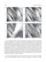
... are 31 0 nm 33 0 nm. (See Color Plate.) Fig. 3 AFM imaging of laminin molecules in air shows submolecular structure in the laminin arms (top row). In the sequential images, a single laminin molecule ... aldehyde groups. Cells for live imaging are washed again in PBS containing 10 mg/ml FSG. The scanning experiments are carried out in culture medium for living cells and in PBS f...
Ngày tải lên: 06/08/2014, 02:20

Atomic Force Microscopy in Cell Biology Episode 2 Part 4 pdf
... by atomic force microscopy. FEBS Lett. 38 1, 161–164. Mou, J., Sheng, S., Ho, R., and Shao, Z. (1996). Chaperonins GroEL and GroES: views form atomic force microscopy. Biophys. J. 71, 22 13 22 21. Mou, ... microtiter wells by scanning force microscopy. Langmuir 11, 1 822 –1 826 . Rogers, W., and Glaser, M. (19 93) . Distributions of proteins and lipids in erythrocyte membrane...
Ngày tải lên: 06/08/2014, 02:20
Atomic Force Microscopy in Cell Biology Episode 2 Part 5 pptx
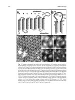
... crystal. 13. Atomic Force Microscopy and Spectroscopy of Membrane Proteins 27 9 Fig. 14 Native bacteriorhodopsin assembled into an orthorhombic lattice. (A) In this crystal form (p 22 1 2 1 ) the ... native 13. Atomic Force Microscopy and Spectroscopy of Membrane Proteins 27 1 Fig. 8 Contact mode AFM topograph of α-hemolysin oligomers. The α-hemolysin inserted into the ph...
Ngày tải lên: 06/08/2014, 02:20